Copyright
©The Author(s) 2019.
World J Orthop. Jun 18, 2019; 10(6): 255-261
Published online Jun 18, 2019. doi: 10.5312/wjo.v10.i6.255
Published online Jun 18, 2019. doi: 10.5312/wjo.v10.i6.255
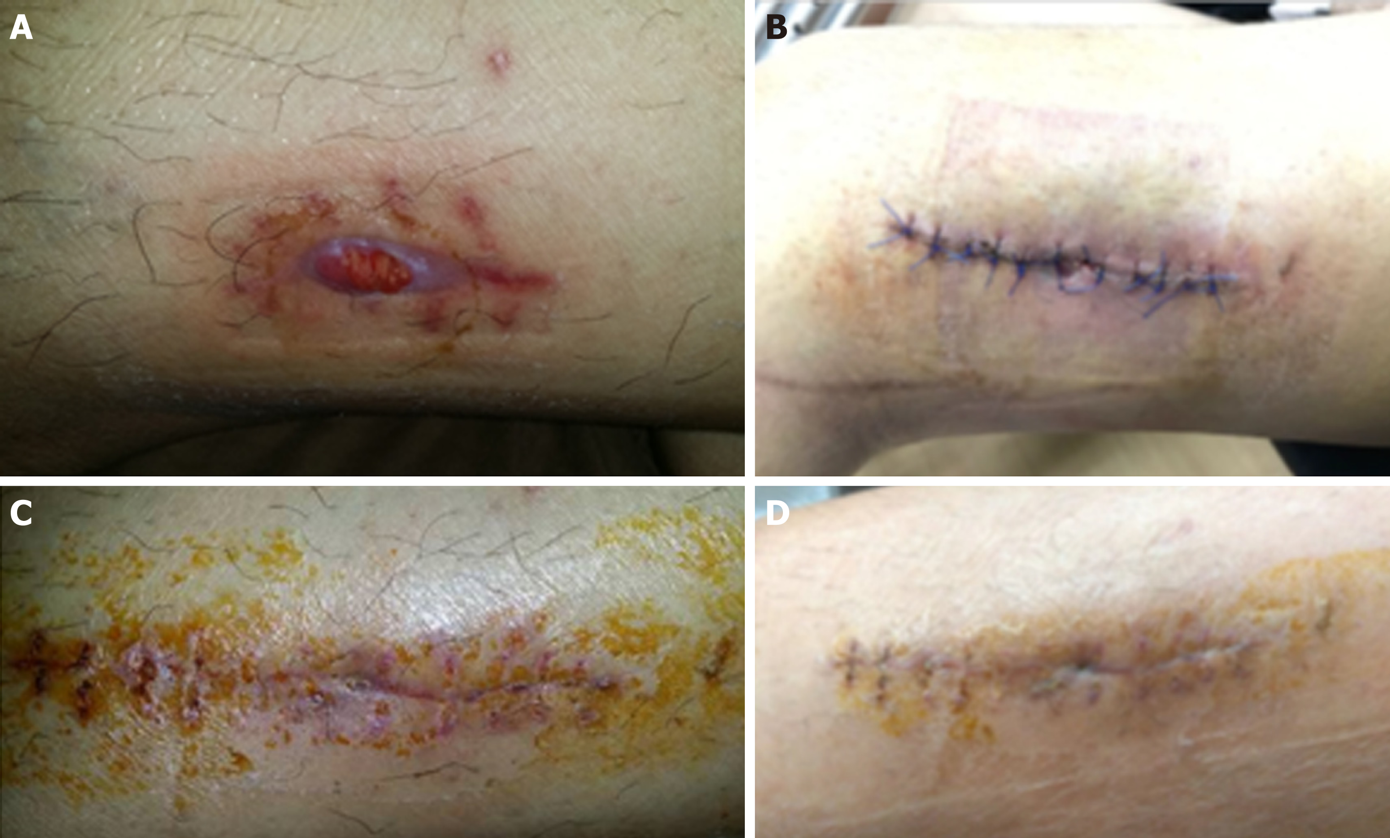
Figure 1 Wound site throughout recovery.
A: Showing pre-operative discharging sinus; B: Post-operative wound showing wound dehiscence and discharge; C: Wound at six weeks after commencing intra-venous Ertapenem; D: Well healed wound.
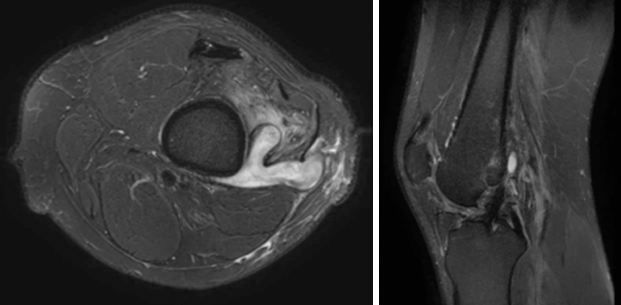
Figure 2 Magnetic resonance imaging knee showing a fistulous communication of a complex Y-shaped abscess in the lateral aspect of the distal thigh extending towards the femoral tunnel of the anterior cruciate ligament reconstruction.
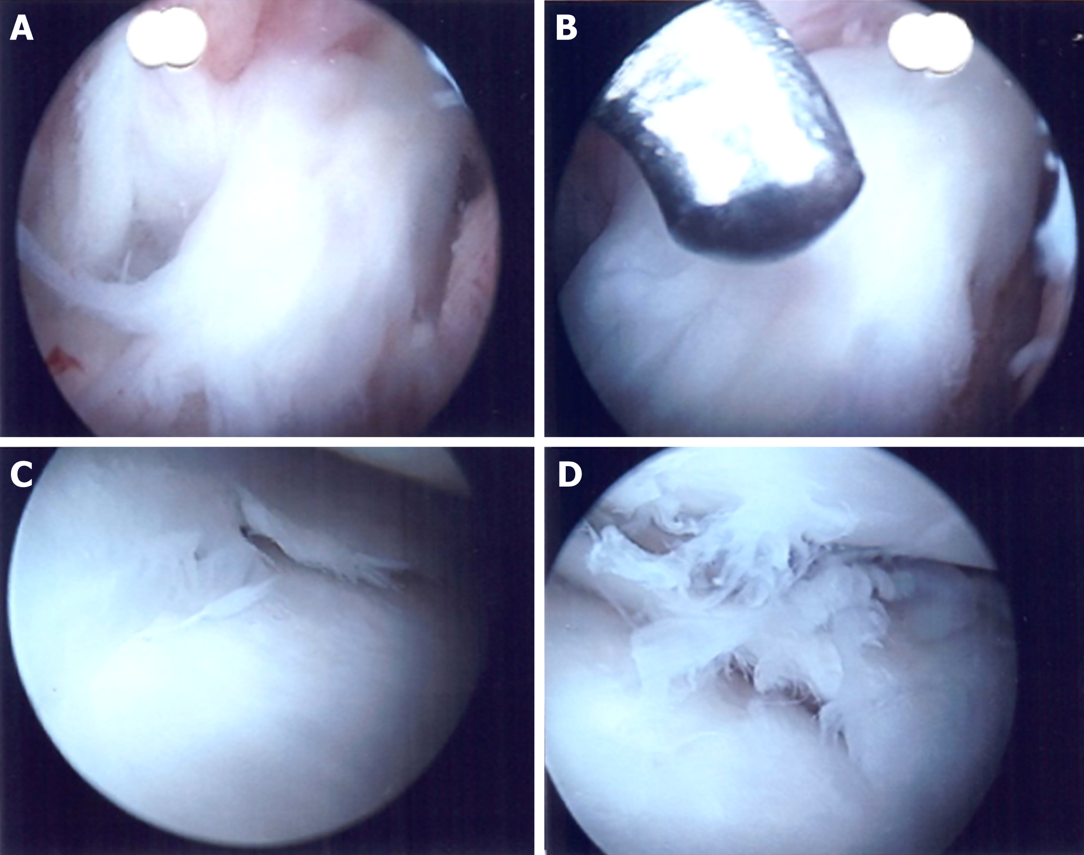
Figure 3 Arthroscopic images of the left knee joint.
No evidence of purulent joint fluid. A, B: Anterior cruciate ligament graft was noted to be intact and healthy; C, D: Partial oblique tear of the medial meniscus as well as degenerative fraying of the lateral meniscus were also noted.
- Citation: Koh D, Tan SM, Tan AHC. Recurrent surgical site infection after anterior cruciate ligament reconstruction: A case report. World J Orthop 2019; 10(6): 255-261
- URL: https://www.wjgnet.com/2218-5836/full/v10/i6/255.htm
- DOI: https://dx.doi.org/10.5312/wjo.v10.i6.255


