Copyright
©The Author(s) 2019.
World J Orthop. Apr 18, 2019; 10(4): 212-218
Published online Apr 18, 2019. doi: 10.5312/wjo.v10.i4.212
Published online Apr 18, 2019. doi: 10.5312/wjo.v10.i4.212
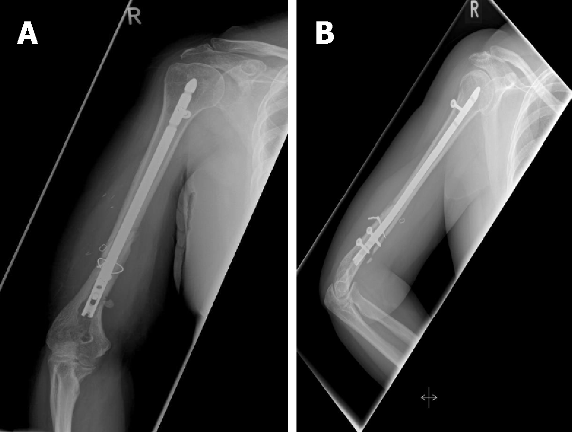
Figure 1 Implant failure after intramedullary nailing.
A: AP view of the right humerus after retrograde intramedullary nailing and implant failure; B: Lateral view with displaced distal fragment.
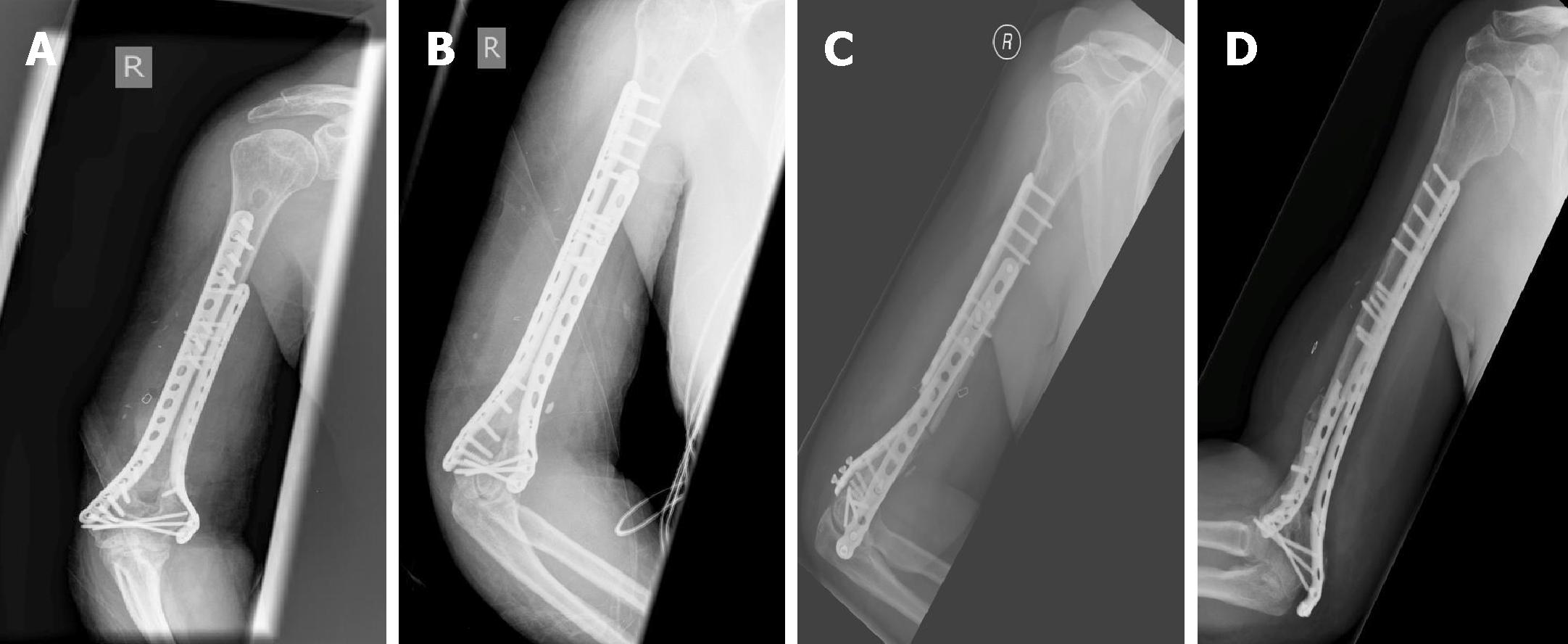
Figure 2 Second implant failure after ORIF.
A: AP view of the right humerus shortly after revision using double plate fixation; B: Lateral view; C: AP view 14 mo after revision to ORIF; D: Lateral view showing implant failure.
Figure 3 Induced membrane technique.
A: Intraoperative picture of the cement application to the humeral defect site after implantation of two nancy nails; B: Hardened cement used as a temporary space holder; C, D: AP and lateral X-ray after first surgery and implantation of two nancy nails.
Figure 4 Vascularized fibula graft and plate fixation.
A: Vascularized fibula graft (VFG) after harvesting. B: Final plate fixation for stabilization of the VFG and bridging the defect. C, D: Lateral and AP X-ray after double plate fixation and combined vascularized fibular graft.
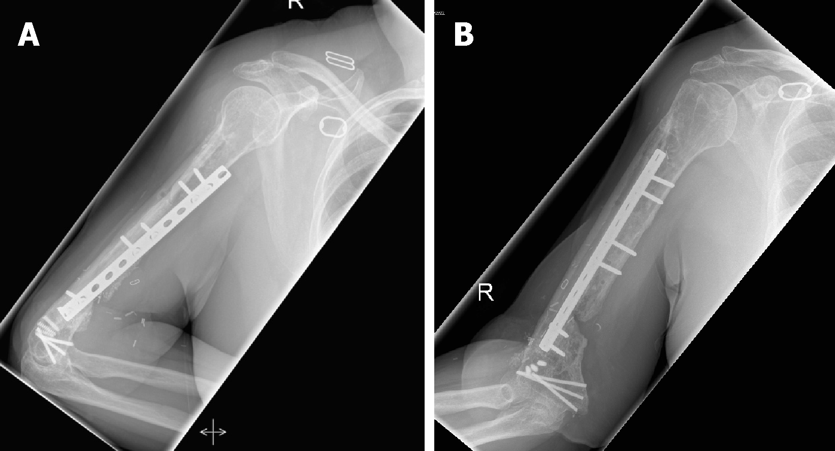
Figure 5 Final result.
A: Final lateral X-ray after removal of one plate and osseous integration of the VFG; B: AP view with a good sight on the integrated VFG. VFG: Vascularized fibula graft.
- Citation: Gathen M, Norris G, Kay S, Giannoudis PV. Recalcitrant distal humeral non-union following previous Leiomyosarcoma excision treated with retainment of a radiated non-angiogenic segment augmented with 20 cm free fibula composite graft: A case report. World J Orthop 2019; 10(4): 212-218
- URL: https://www.wjgnet.com/2218-5836/full/v10/i4/212.htm
- DOI: https://dx.doi.org/10.5312/wjo.v10.i4.212


