Copyright
©The Author(s) 2019.
World J Orthop. Feb 18, 2019; 10(2): 115-122
Published online Feb 18, 2019. doi: 10.5312/wjo.v10.i2.115
Published online Feb 18, 2019. doi: 10.5312/wjo.v10.i2.115
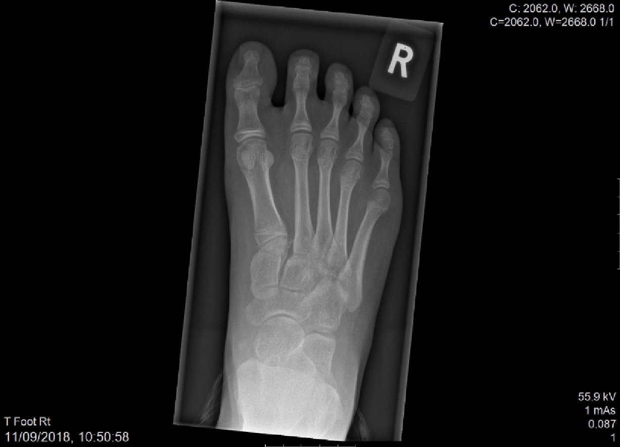
Figure 1 Pre-operative X-ray showing increased distance between 1st and 2nd metatarsals with small bone fragment (inside circle).
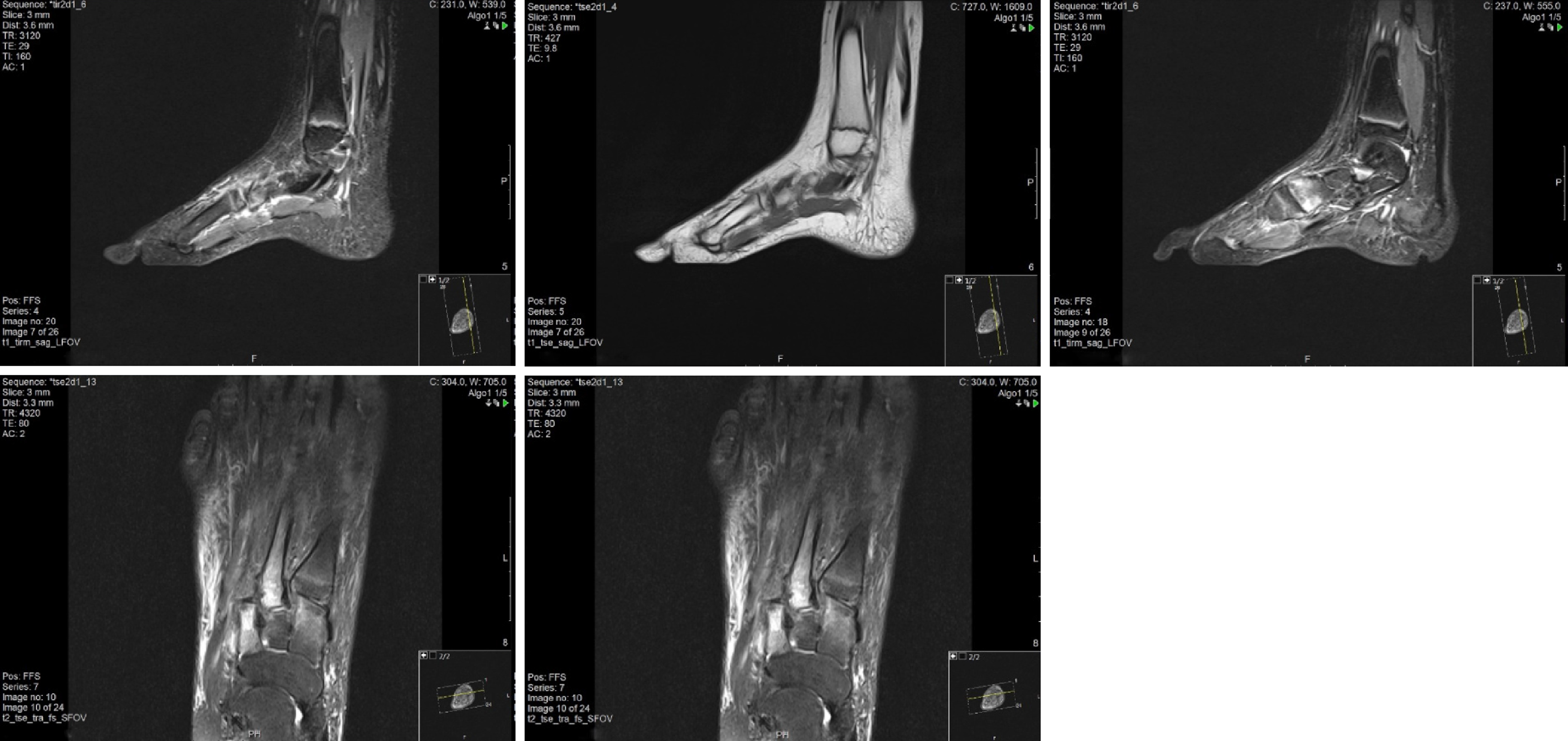
Figure 2 Magnetic resonance imaging image series.
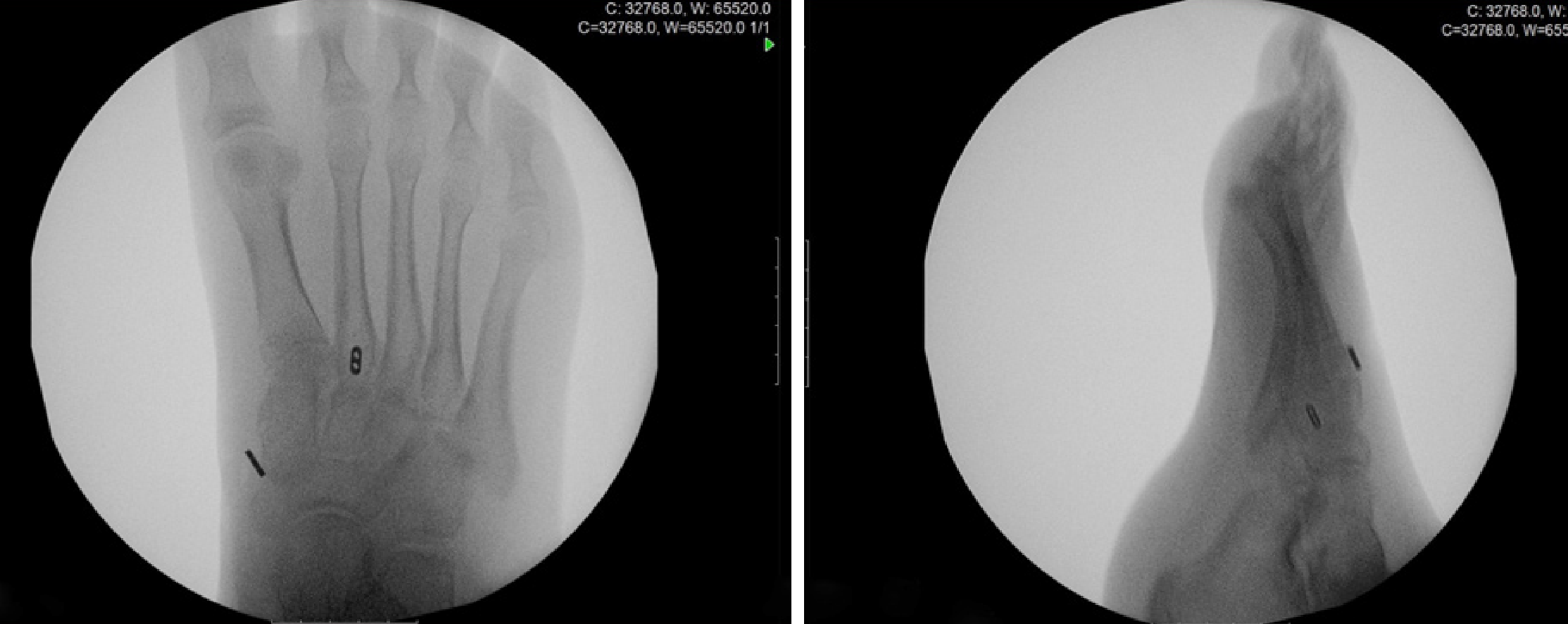
Figure 3 Intra-operative X-ray confirmed maintance of reduction after removal of bone reduction clamp.
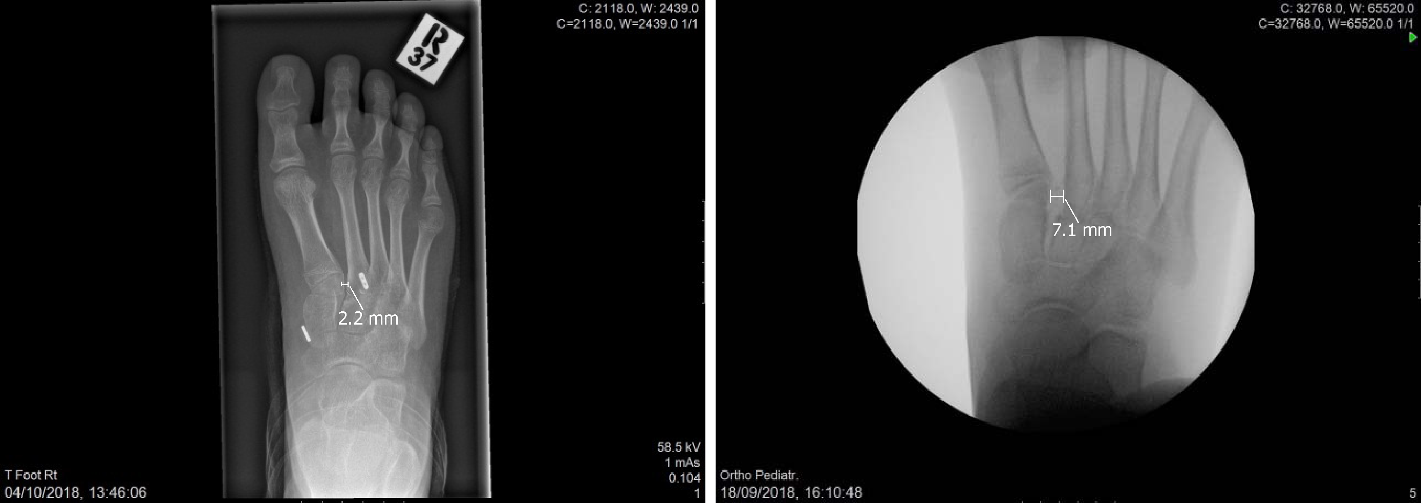
Figure 4 Post-operative x-ray (3rd week) on the left side and pre-operative X-ray on the right side.
- Citation: Tzatzairis T, Firth G, Parker L. Adolescent Lisfranc injury treated with TightRopeTM: A case report and review of literature. World J Orthop 2019; 10(2): 115-122
- URL: https://www.wjgnet.com/2218-5836/full/v10/i2/115.htm
- DOI: https://dx.doi.org/10.5312/wjo.v10.i2.115


