Copyright
©The Author(s) 2019.
World J Orthop. Oct 18, 2019; 10(10): 364-370
Published online Oct 18, 2019. doi: 10.5312/wjo.v10.i10.364
Published online Oct 18, 2019. doi: 10.5312/wjo.v10.i10.364
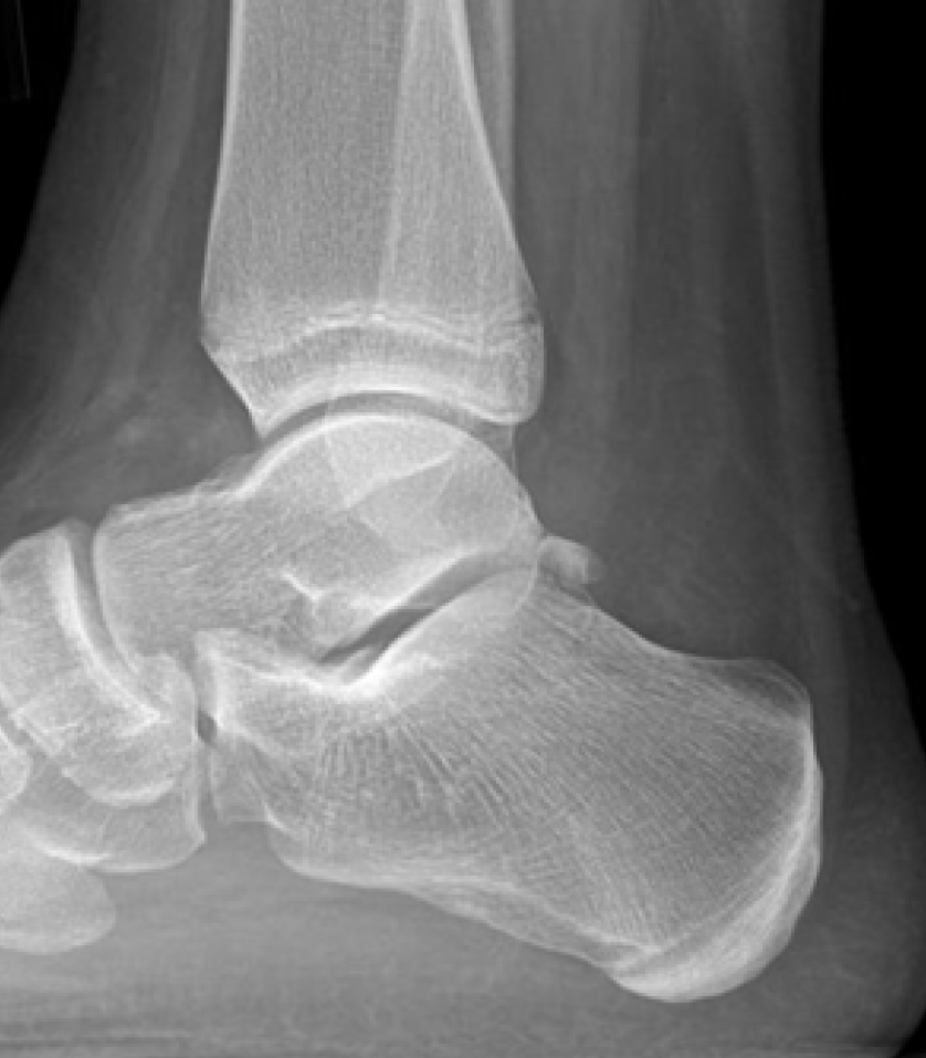
Figure 1 Fifteen-year old male with posterior ankle pain with os trigonum seen on lateral ankle radiograph.
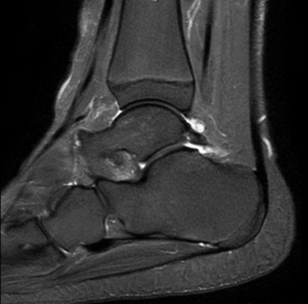
Figure 2 Magnetic resonance imaging-sagittal image demonstrating edema-like signal intensity adjacent to the os trigonum in the previously mentioned 15-year-old patient in Figure 1.
- Citation: Kushare I, Kastan K, Allahabadi S. Posterior ankle impingement–an underdiagnosed cause of ankle pain in pediatric patients. World J Orthop 2019; 10(10): 364-370
- URL: https://www.wjgnet.com/2218-5836/full/v10/i10/364.htm
- DOI: https://dx.doi.org/10.5312/wjo.v10.i10.364


