Copyright
©The Author(s) 2025.
World J Clin Oncol. Aug 24, 2025; 16(8): 108865
Published online Aug 24, 2025. doi: 10.5306/wjco.v16.i8.108865
Published online Aug 24, 2025. doi: 10.5306/wjco.v16.i8.108865
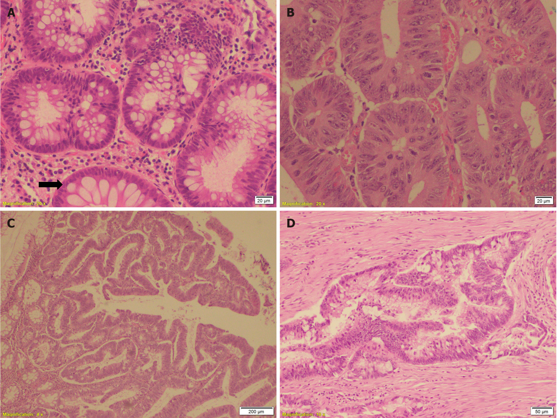
Figure 1 Progression of adenoma to colon cancer.
A: Tubular adenomas with low-grade dysplasia showing dysplastic epithelium with elongated and hyperchromatic nuclei and variable amounts of mucin. Note abrupt transition from normal (arrow) to dysplastic mucosa [hematoxylin and eosin (HE) × 20]; B: Tubular adenomas with high-grade dysplasia showing cribriform pattern, nuclear stratification through the whole thickness of the epithelium, prominent nucleoli, loss of cell polarity, marked pleomorphism and atypical mitosis (HE × 20); C: Tubulovillous adenoma exhibiting admixture of tubular and villous components (HE × 4); D: Moderately differentiated infiltrating adenocarcinoma throughout the wall of colon (HE × 10).
- Citation: Esheba GE, Kamel HF, Youssef HM, Badawood HF, Alshamrani AA, Alharbi RJ, Nassir R. Mutational profile of a Saudi patient with Familial adenomatous polyposis that progressed to colon cancer: A case report. World J Clin Oncol 2025; 16(8): 108865
- URL: https://www.wjgnet.com/2218-4333/full/v16/i8/108865.htm
- DOI: https://dx.doi.org/10.5306/wjco.v16.i8.108865


