Copyright
©The Author(s) 2024.
World J Clin Oncol. Feb 24, 2024; 15(2): 317-328
Published online Feb 24, 2024. doi: 10.5306/wjco.v15.i2.317
Published online Feb 24, 2024. doi: 10.5306/wjco.v15.i2.317
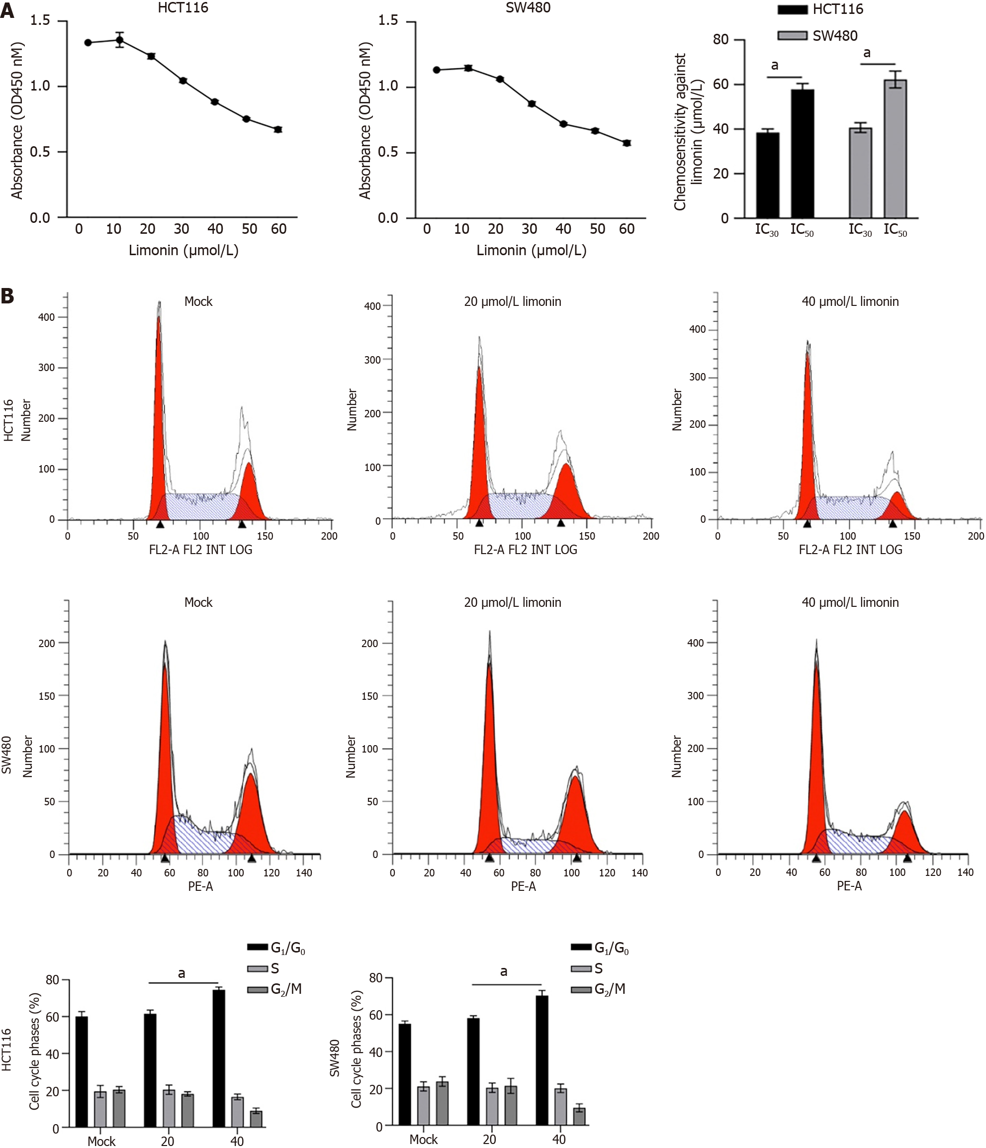
Figure 1 Limonin inhibits cell viability and cell cycle distribution in colorectal cancer cells.
A: After being cultured with 10-60 μmol/L limonin for 24 h, cell viability was measured by performing cholecystokinin octapeptide-8 and IC30 or IC50 was calculated; B: After being cultured with 20 or 40 μmol/L limonin for 24 h, cell cycle distribution was measured. aP < 0.05 vs Mock group.
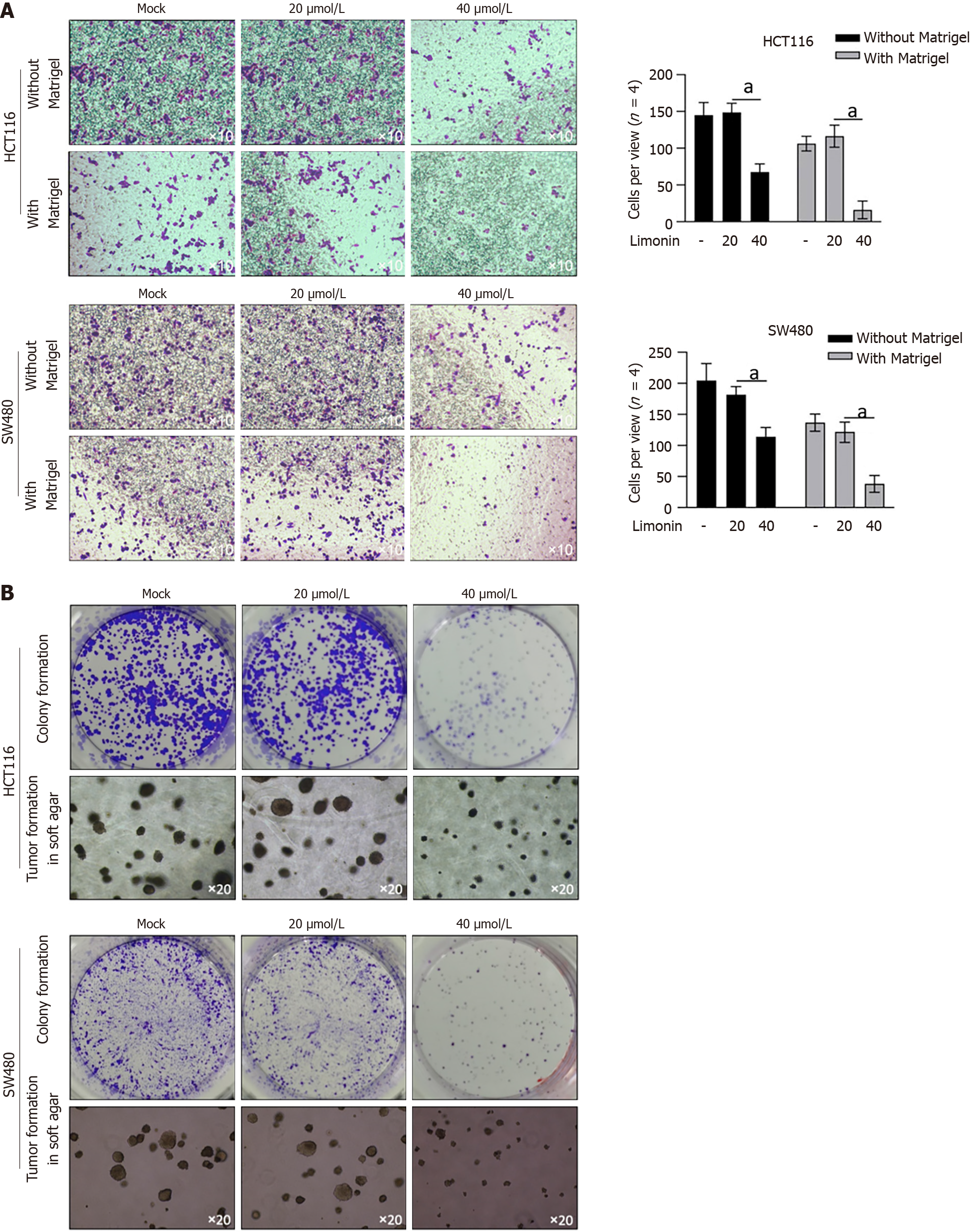
Figure 2 Limonin inhibits malignancies in colorectal cancer cells.
A: After being cultured with 20 or 40 μmol/L limonin for 24 h without Matrigel, or for 48 h with Matrigel, cells transferred to lower membrane were fixed, stained and counted. aP < 0.05 vs Mock group; B: After being cultured with 20 or 40 μmol/L limonin for 14 d, colonies on plates or soft agar were fixed and stained.
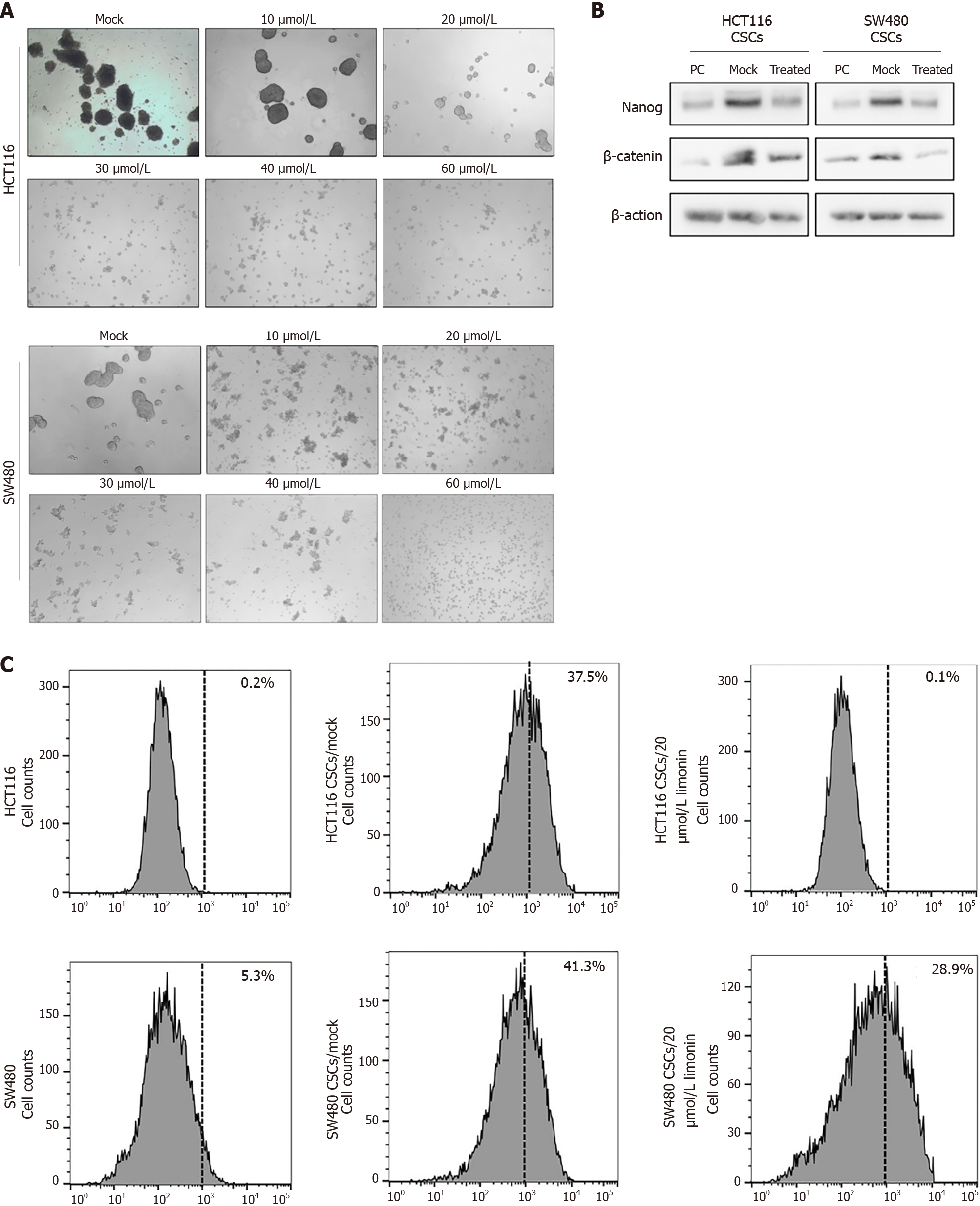
Figure 3 Limonin inhibits stemness in cancer stem-like cells derived from colorectal cancer cells.
A: After being culture with 10-60 μmol/L limonin for 10 d in serum-free medium, spheroid formation was observed; B: 20 μmol/L limonin was employed for treatment, and stemness hallmarkers, including Nanog and β-catenin were measured by western blot; C: Aldehyde dehydrogenase 1-positive proportion was measured by flow cytometry. PC: Parental cells; CSCs: Cancer stem-like cells.
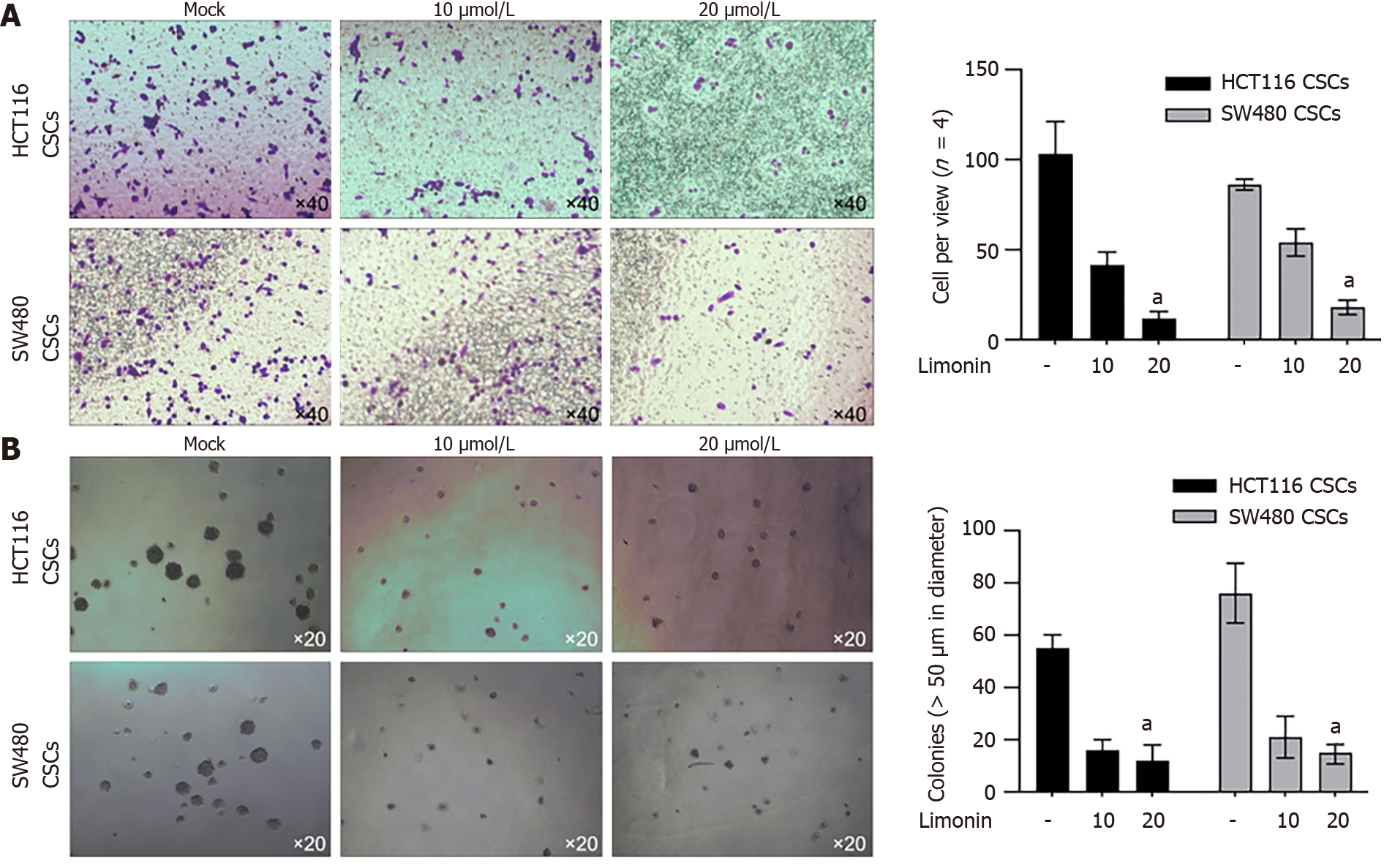
Figure 4 Limonin inhibits malignancies in cancer stem-like cells derived from colorectal cancer cells.
A: Cell invasion capacity was measured by performing transwell assay after being cultured with 10 or 20 μmol/L limonin; B: After limonin treatment, tumor formation in soft agar was performed. aP < 0.05 vs Mock group. CSCs: Cancer stem-like cells.
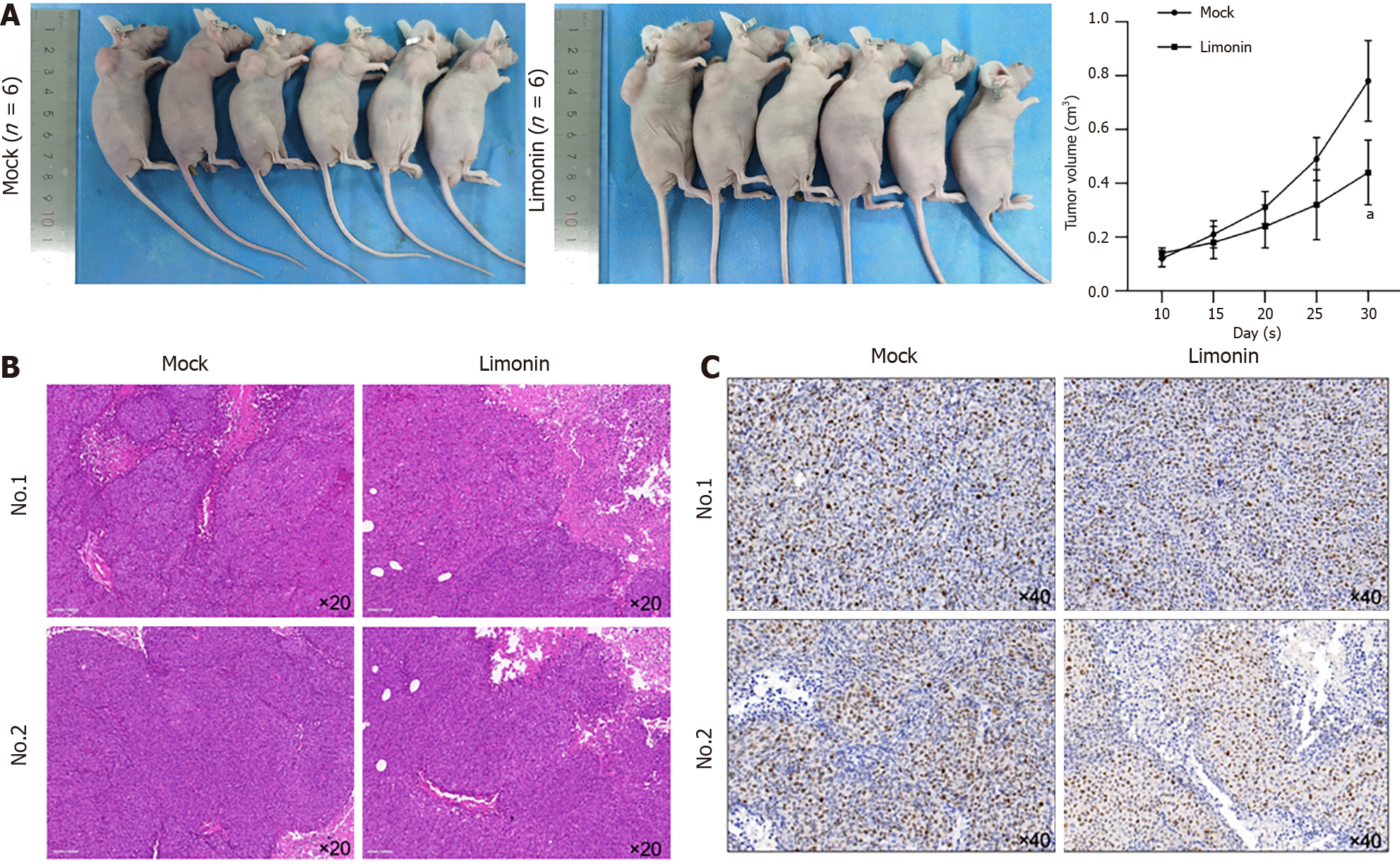
Figure 5 Limonin pre-treatment inhibits tumor formation in nude mice.
A: Cancer stem-like cells pretreated using 20 μmol/L limonin or not were seeded in nude mice and be maintained for 30 d. From day 10, tumor size was measured every five days. aP < 0.05 vs Mock group; B: Hematoxylin and eosin staining was performed to detect physiological morphology; C: Ki67 was stained.
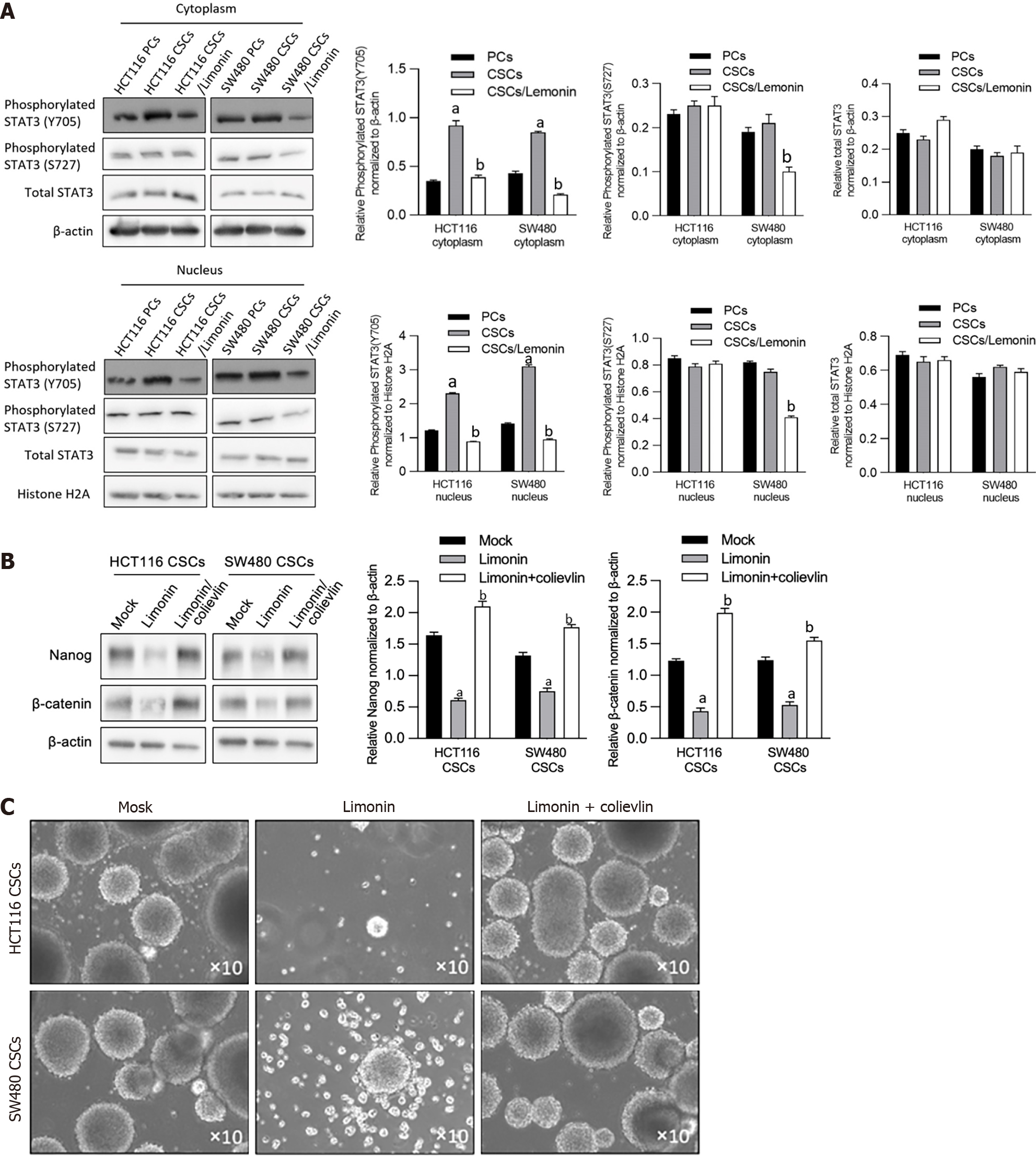
Figure 6 Limonin inhibits STAT3 signaling.
A: After being cultured using limonin, total STAT3, phosphorylated STAT3 (Y705) or STAT3 (S727), in cytoplasmic or nucleus fraction, were measured by performing western blot; B: Stemness hallmarkers, including Nanog and β-catenin, were detected by western blot after Limonin treatment with or without 2.5 μmol/L colievlin pretreatment; C: Sphere formation was observed after Limonin treatment with or without 2.5 μmol/L colievlin pretreatment. aP < 0.05 vs parental cells group; bP < 0.05 vs cancer stem-like cells group. PCs: Parental cells; CSCs: Cancer stem-like cells.
- Citation: Zhang WF, Ruan CW, Wu JB, Wu GL, Wang XG, Chen HJ. Limonin inhibits the stemness of cancer stem-like cells derived from colorectal carcinoma cells potentially via blocking STAT3 signaling. World J Clin Oncol 2024; 15(2): 317-328
- URL: https://www.wjgnet.com/2218-4333/full/v15/i2/317.htm
- DOI: https://dx.doi.org/10.5306/wjco.v15.i2.317


