Copyright
©2011 Baishideng Publishing Group Co.
World J Gastrointest Pathophysiol. Feb 15, 2011; 2(1): 15-18
Published online Feb 15, 2011. doi: 10.4291/wjgp.v2.i1.15
Published online Feb 15, 2011. doi: 10.4291/wjgp.v2.i1.15
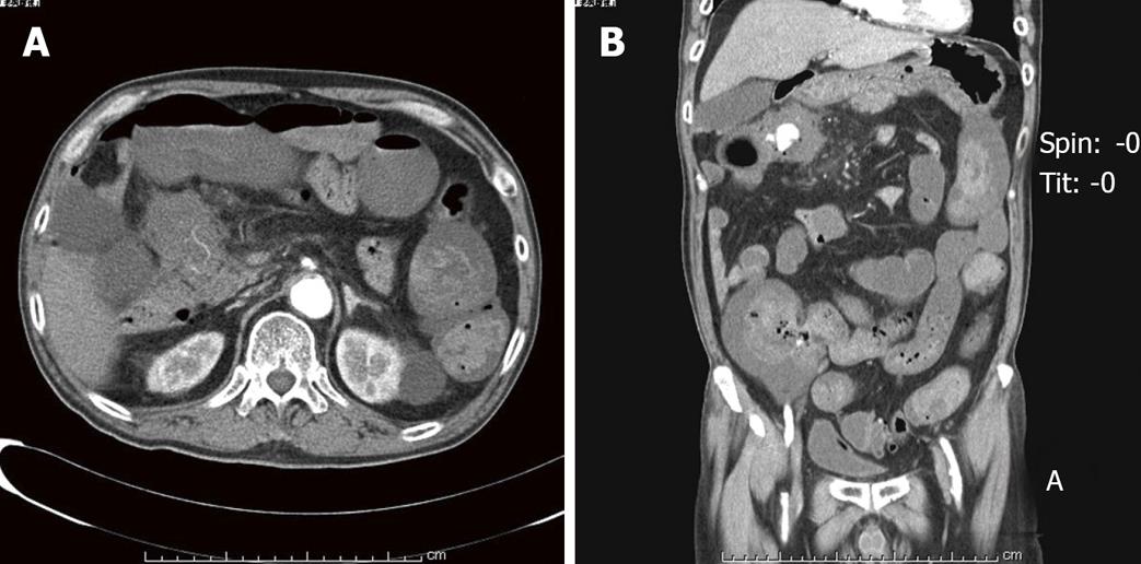
Figure 1 A abdominal computed tomography scan.
A: The tumor invading the pancreas and duodenum; B: Extravasation of blood from the gastroduodenal artery into the ascending colon.
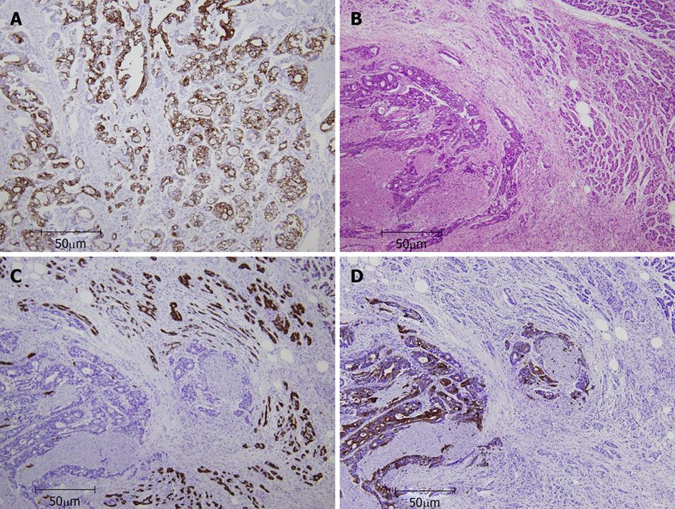
Figure 2 Immunohistochemical staining of cytokeratin 7 and CK20.
A: Colonic tumor cells were cytoplasm-positive for cytokeratin (CK)20; B: H&E staining shows the border between pancreatic tumor and normal pancreas; C: Pancreatic tumor cells are cytoplasm-positive for CK20 but not for CK7; D:Immunohistochemical staining of normal pancreatic tissue shows positivity for CK7 but negativity for CK20. These CK7/CK20 immunostaining images were obtained for the same field.
- Citation: Iwata T, Konishi K, Yamazaki T, Kitamura K, Katagiri A, Muramoto T, Kubota Y, Yano Y, Kobayashi Y, Yamochi T, Ohike N, Murakami M, Gokan T, Yoshikawa N, Imawari M. Right colon cancer presenting as hemorrhagic shock. World J Gastrointest Pathophysiol 2011; 2(1): 15-18
- URL: https://www.wjgnet.com/2150-5330/full/v2/i1/15.htm
- DOI: https://dx.doi.org/10.4291/wjgp.v2.i1.15


