Copyright
©The Author(s) 2025.
World J Gastrointest Pathophysiol. Jun 22, 2025; 16(2): 107599
Published online Jun 22, 2025. doi: 10.4291/wjgp.v16.i2.107599
Published online Jun 22, 2025. doi: 10.4291/wjgp.v16.i2.107599
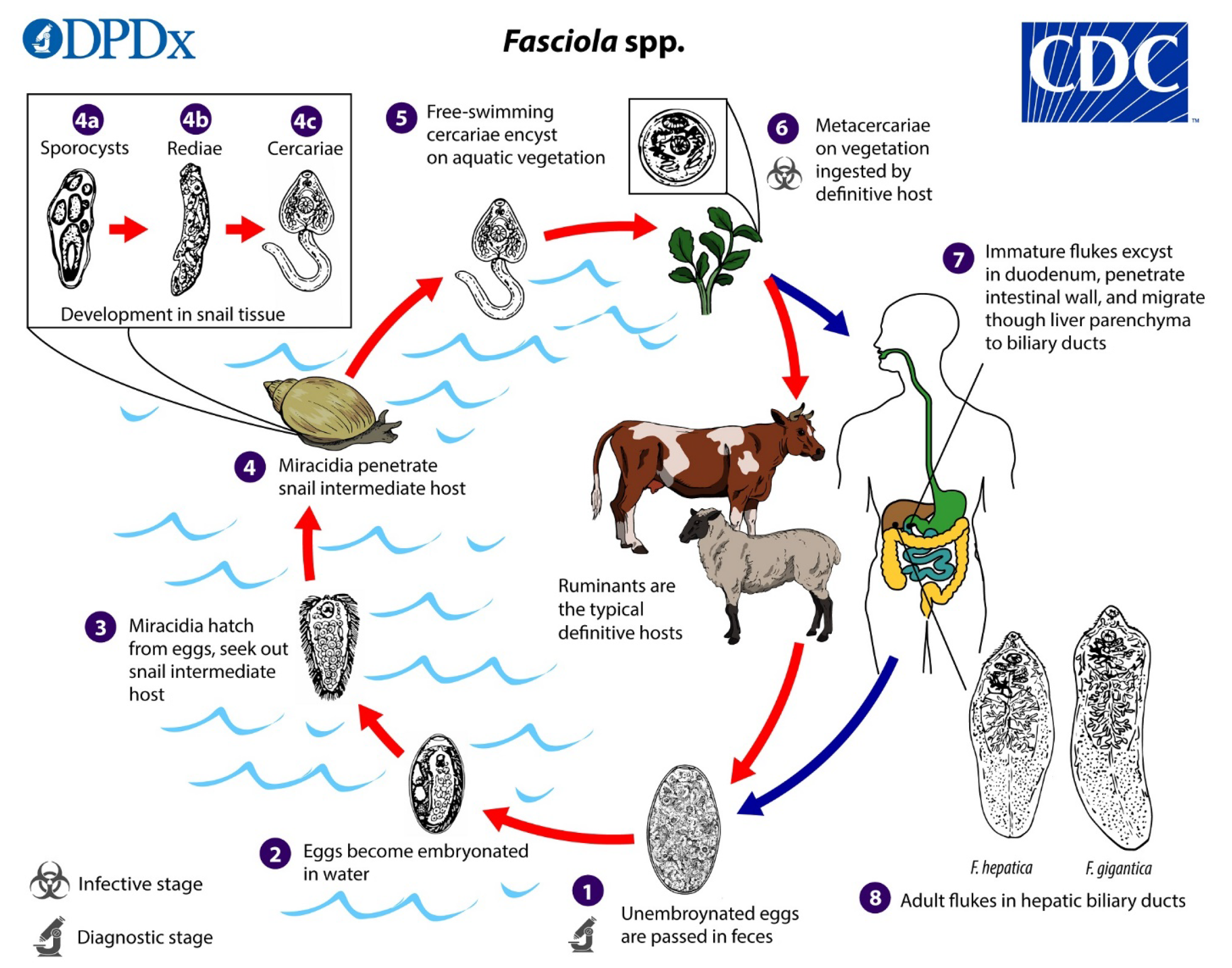
Figure 1 Life cycle of Fasciola spp.
Immature eggs are discharged in the biliary ducts and passed in the stool (1). Eggs become embryonated in freshwater over appro
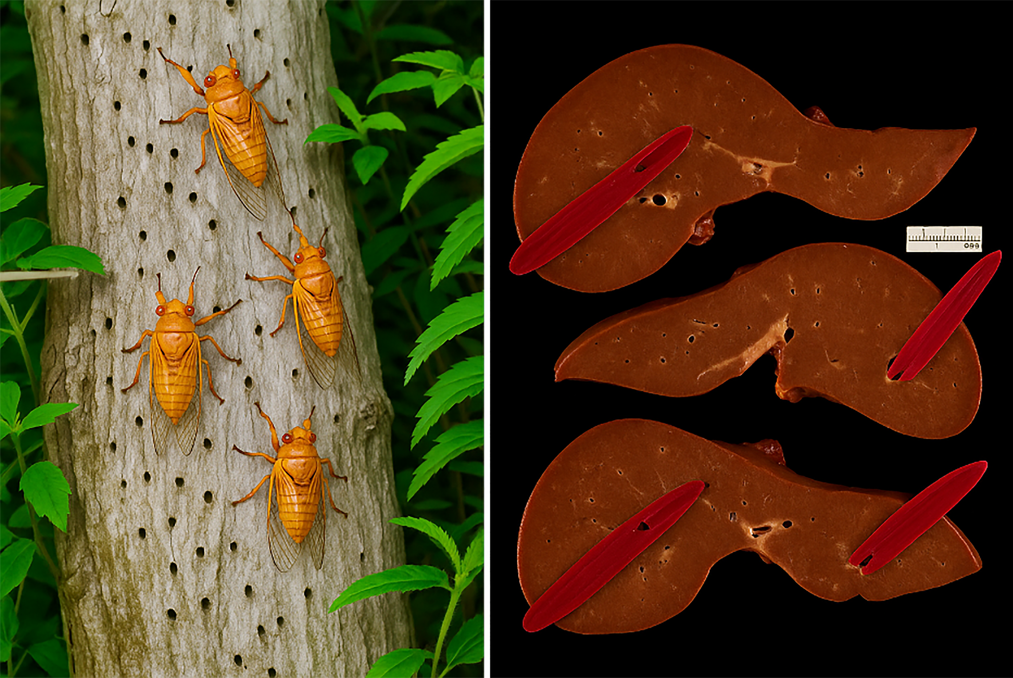
Figure 2 The termite's analogy illustration.
We suggest calling the liver flukes the liver “termites” emphasizing the inhabitation and damage to the biliary tract and hepatic parenchyma.
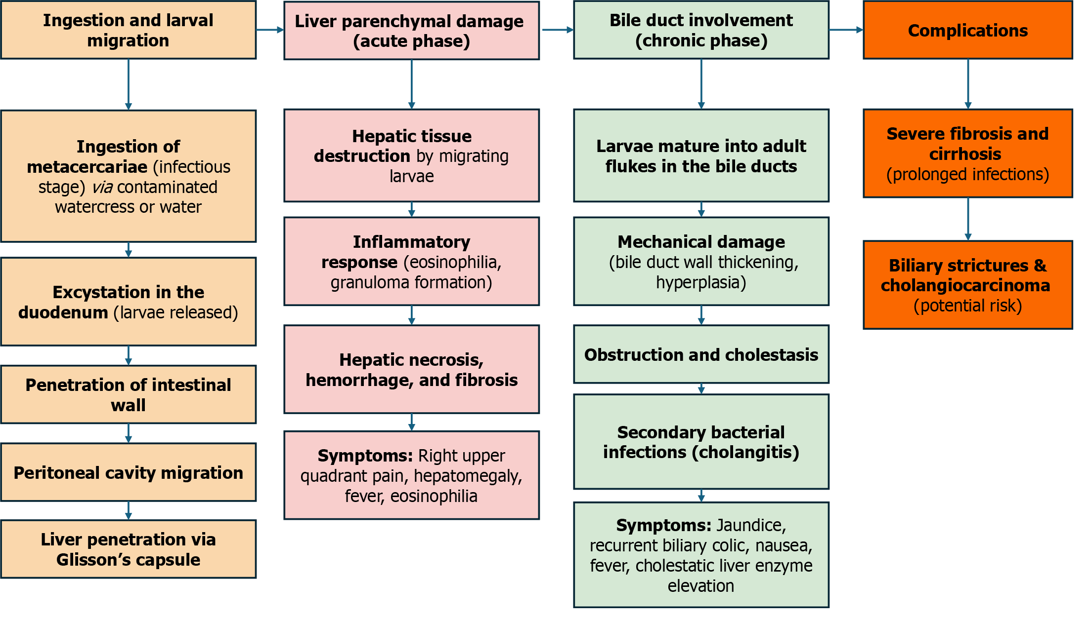
Figure 3
Flowchart of mechanisms of liver and bile duct involvement and how they lead to symptoms.
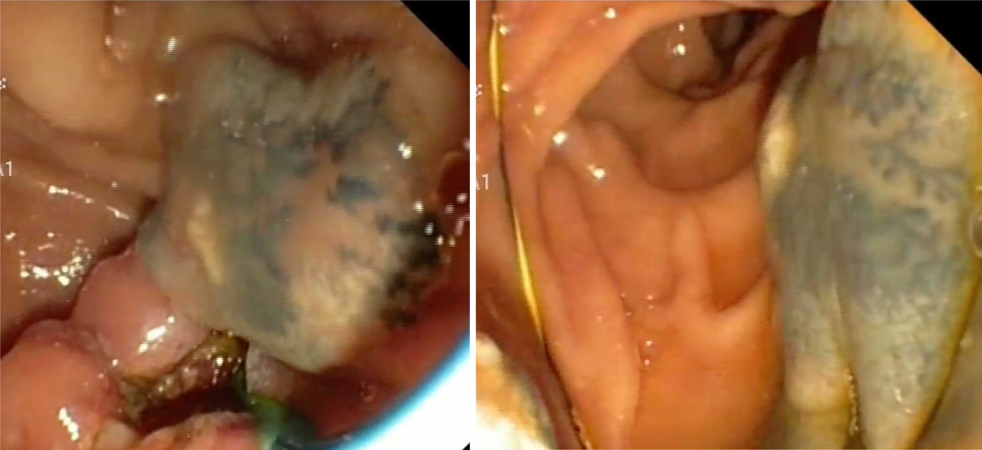
Figure 4
Viable Fasciola spp.
Specimens retrieved from the gallbladder during endoscopic retrograde cholangiopancreatography.
- Citation: Ismail A, Abdelsalam MA, Shahin MH, Ahmed Y, Bahcecioglu IH, Yalniz M, Tawheed A. Hepatobiliary fascioliasis: A neglected re-emerging threat, its diagnostic and management challenges. World J Gastrointest Pathophysiol 2025; 16(2): 107599
- URL: https://www.wjgnet.com/2150-5330/full/v16/i2/107599.htm
- DOI: https://dx.doi.org/10.4291/wjgp.v16.i2.107599


