Copyright
©The Author(s) 2019.
World J Gastrointest Pathophysiol. Sep 10, 2019; 10(2): 29-35
Published online Sep 10, 2019. doi: 10.4291/wjgp.v10.i2.29
Published online Sep 10, 2019. doi: 10.4291/wjgp.v10.i2.29
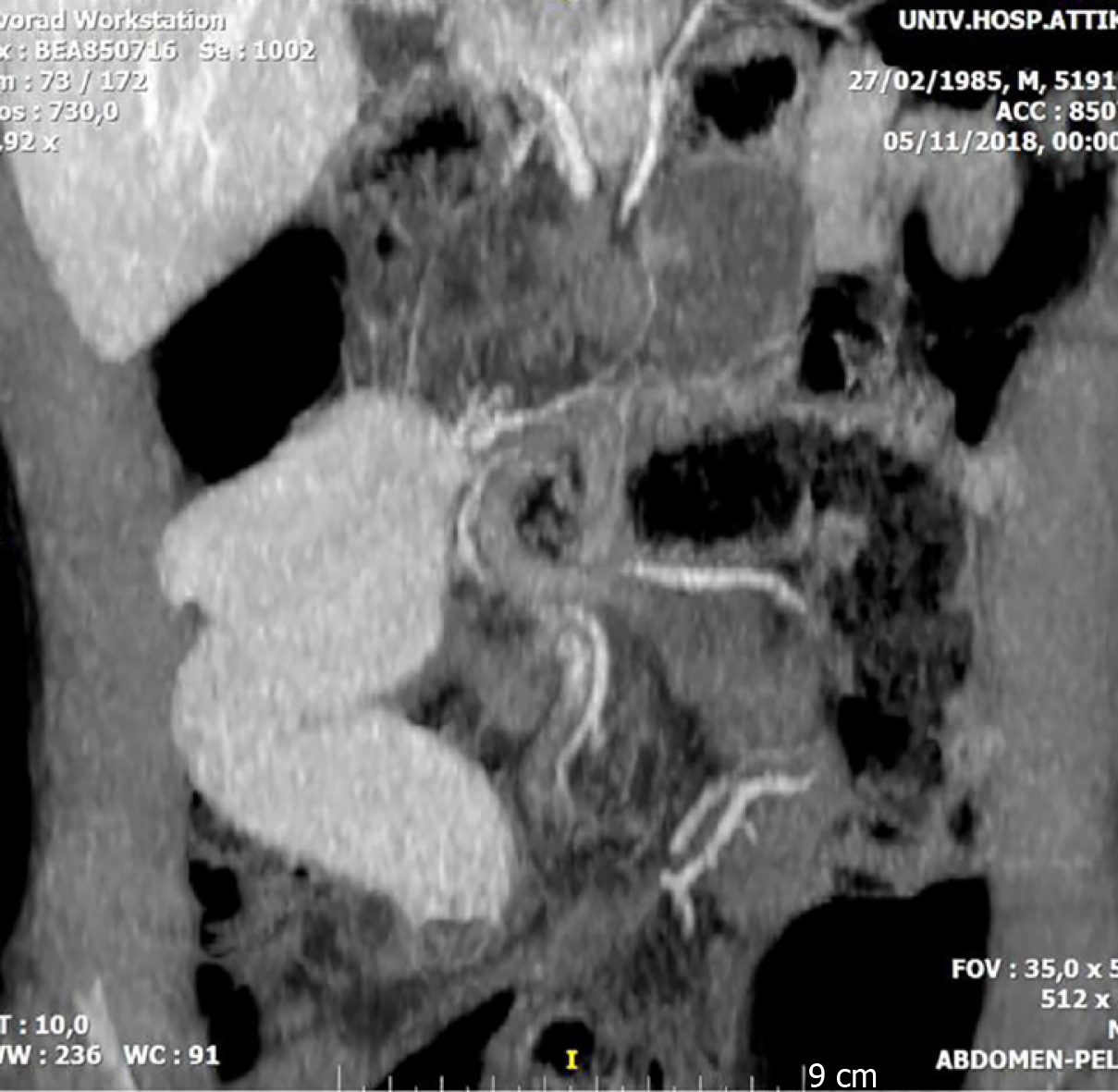
Figure 1 Abdominal computed tomography indicative of the torsion of the ileum around the adhesion at the center of the picture.
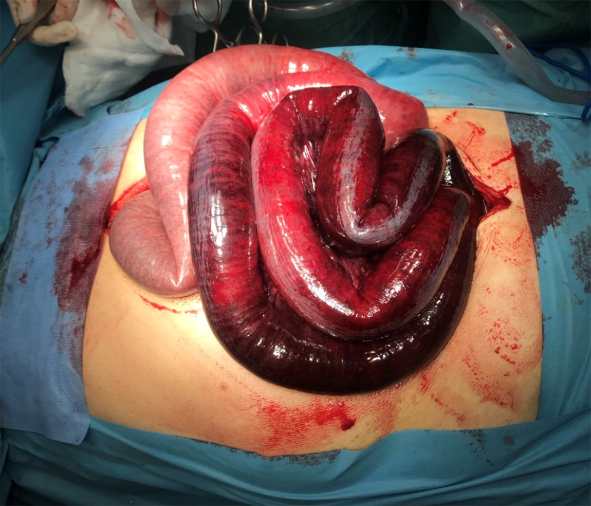
Figure 2 Dilated ischemic small bowel loops at the exploratory laparotomy.
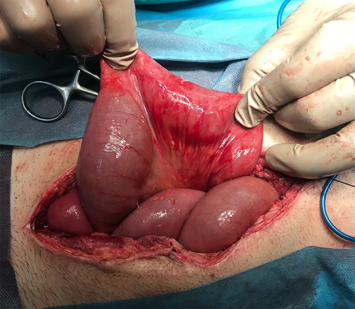
Figure 3 The small bowel at second look laparotomy 48 h later.
Location of most severe ischemia: On the left side of the image the surgeon holds the bowel at the location of previous anastomosis were the adhesion that lead to torsion and ischemia was formed.
- Citation: Vassiliu P, Ntella V, Theodoroleas G, Mantanis Z, Pentara I, Papoutsi E, Mastoraki A, Arkadopoulos N. Successful management of adhesion related small bowel ischemia without intestinal resection: A case report and review of literature. World J Gastrointest Pathophysiol 2019; 10(2): 29-35
- URL: https://www.wjgnet.com/2150-5330/full/v10/i2/29.htm
- DOI: https://dx.doi.org/10.4291/wjgp.v10.i2.29


