Copyright
©The Author(s) 2019.
World J Gastrointest Pathophysiol. Sep 10, 2019; 10(2): 17-28
Published online Sep 10, 2019. doi: 10.4291/wjgp.v10.i2.17
Published online Sep 10, 2019. doi: 10.4291/wjgp.v10.i2.17
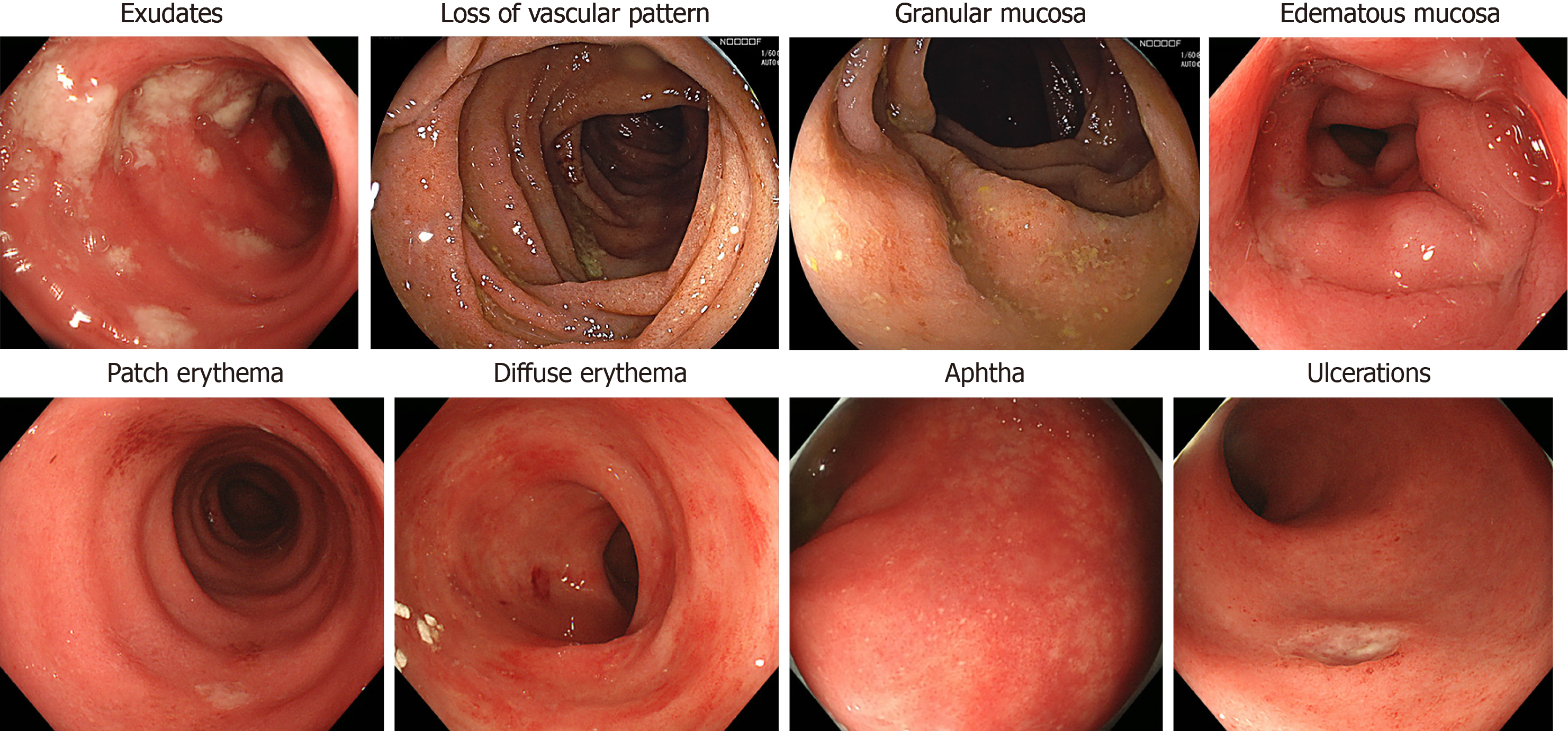
Figure 1 Endoscopic findings caused by an immune checkpoint inhibitor.
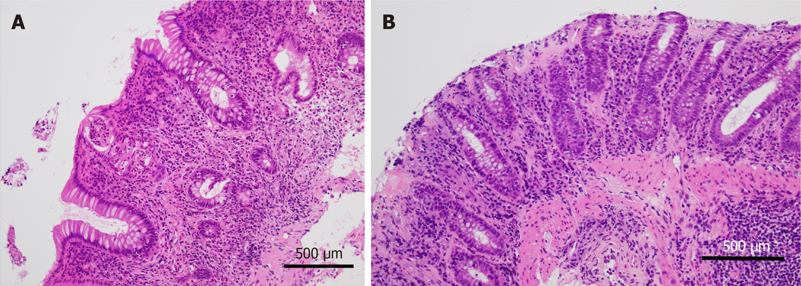
Figure 2 Programmed cell death protein 1 inhibitor-associated colitis.
A: This colon biopsy reveals lamina propria expansion by lymphoplasmacytic infiltrate. Crypt distortion, crypt abscess, and cryptitis are prominent in the mucosa. In the stroma, a significantly increased eosinophilic infiltrate is observed; B: In another case of immune checkpoint inhibitors-related colitic mucosa, a subluminal collagen band thickening is prominent as observed in collagenous colitis. (Hematoxylin and eosin original magnification × 20, a scale bar represents 500 µm).
- Citation: Nishida T, Iijima H, Adachi S. Immune checkpoint inhibitor-induced diarrhea/colitis: Endoscopic and pathologic findings. World J Gastrointest Pathophysiol 2019; 10(2): 17-28
- URL: https://www.wjgnet.com/2150-5330/full/v10/i2/17.htm
- DOI: https://dx.doi.org/10.4291/wjgp.v10.i2.17


