Copyright
©The Author(s) 2017.
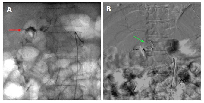
Figure 3 Fluoroscopic images of a case of 2-3 cm bleeding duodenal ulcer that failed endoscopic hemostasis with Endoclip application.
A: Fluoroscopic image following contrast injection to the right gastric artery showing active extravasation into the lumen. The bleeding vessel (red arrow) is identified which is near the endoscopically placed clip; B: Digital subtraction angiography post coiling of the right gastric artery (green arrow).
- Citation: Ray DM, Srinivasan I, Tang SJ, Vilmann AS, Vilmann P, McCowan TC, Patel AM. Complementary roles of interventional radiology and therapeutic endoscopy in gastroenterology. World J Radiol 2017; 9(3): 97-111
- URL: https://www.wjgnet.com/1949-8470/full/v9/i3/97.htm
- DOI: https://dx.doi.org/10.4329/wjr.v9.i3.97


