Copyright
©2014 Baishideng Publishing Group Inc.
World J Radiol. Jun 28, 2014; 6(6): 388-391
Published online Jun 28, 2014. doi: 10.4329/wjr.v6.i6.388
Published online Jun 28, 2014. doi: 10.4329/wjr.v6.i6.388
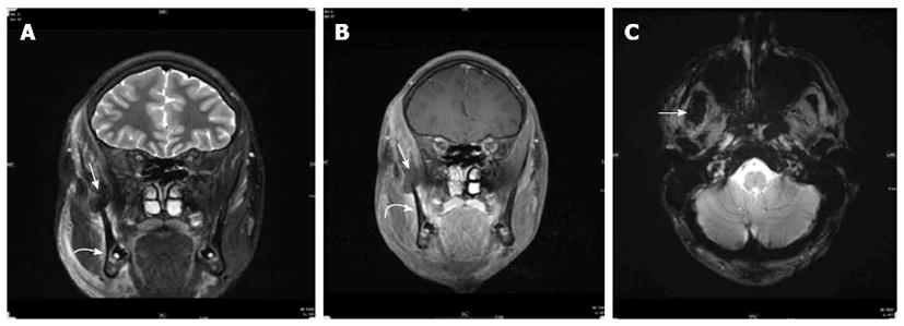
Figure 3 T2 Coronal SPIR (A) and T1 Coronal SPIR post contrast images (B) through the face demonstrate rupture of myotendinous insertion of right temporalis muscle with a 2.
5 cm × 1.5 cm collection surrounding the coronoid region of right mandible at the site of temporalis muscle insertion (straight arrows), the collection demonstrates low signal on T2 weighted imaging (A) and susceptibility artifact/blooming on the T2 axial GRE image (C) consistent with hematoma. Additionally, there is mild swelling, increased T2 signal and mild enhancement in the right masseter muscle notably near its insertion to the anterior mandible as demonstrated on figures A and B suggesting a sprain (curved arrow). Swelling and edema of overlying subcutaneous soft tissues is noted as well.
- Citation: Naffaa LN, Tandon YK, Rubin M. Myotendinous rupture of temporalis muscle: A rare injury following seizure. World J Radiol 2014; 6(6): 388-391
- URL: https://www.wjgnet.com/1949-8470/full/v6/i6/388.htm
- DOI: https://dx.doi.org/10.4329/wjr.v6.i6.388


