Copyright
©2014 Baishideng Publishing Group Inc.
World J Radiol. Oct 28, 2014; 6(10): 808-825
Published online Oct 28, 2014. doi: 10.4329/wjr.v6.i10.808
Published online Oct 28, 2014. doi: 10.4329/wjr.v6.i10.808
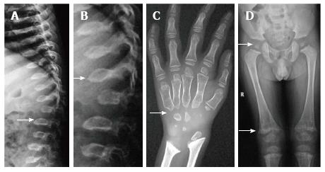
Figure 7 Morquio’s syndrome.
Radiographs of spine (A, B) show platyspondyly with maintained intervertebral disc height (arrow, A) and central beaking (arrow, B). Radiograph of hand (C) shows proximal pointing of metacarpals. Radiograph of pelvis and lower limbs (D) show delayed ossification of femoral heads, irregular epiphyses and secondary metaphyseal widening in proximal femur and around knee joint (arrows).
- Citation: Panda A, Gamanagatti S, Jana M, Gupta AK. Skeletal dysplasias: A radiographic approach and review of common non-lethal skeletal dysplasias. World J Radiol 2014; 6(10): 808-825
- URL: https://www.wjgnet.com/1949-8470/full/v6/i10/808.htm
- DOI: https://dx.doi.org/10.4329/wjr.v6.i10.808


