Copyright
©The Author(s) 2022.
World J Radiol. Jun 28, 2022; 14(6): 137-150
Published online Jun 28, 2022. doi: 10.4329/wjr.v14.i6.137
Published online Jun 28, 2022. doi: 10.4329/wjr.v14.i6.137
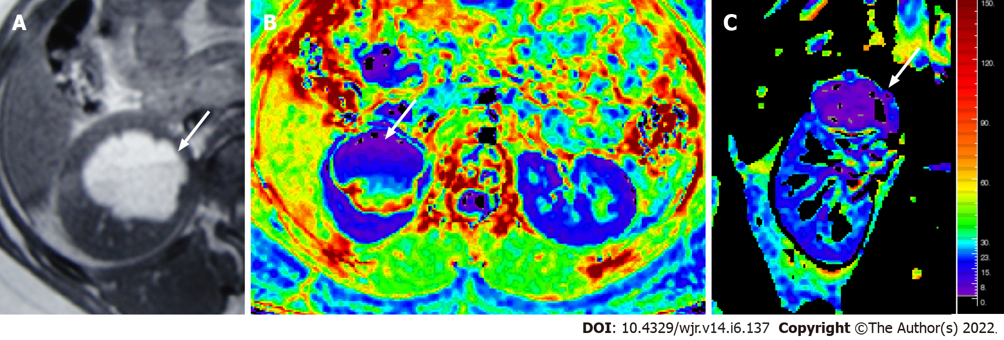
Figure 8 Magnetic resonance images.
T1W axial MR image (A) showing a hyperintense lesion in the upper pole of the right kidney, with fluid-fluid level, suggestive of a hemorrhagic cyst; Axial & coronal R2* maps (B and C) showing an R2* value of 6.1/s.
- Citation: Aggarwal A, Das CJ, Sharma S. Recent advances in imaging techniques of renal masses. World J Radiol 2022; 14(6): 137-150
- URL: https://www.wjgnet.com/1949-8470/full/v14/i6/137.htm
- DOI: https://dx.doi.org/10.4329/wjr.v14.i6.137


