Copyright
©The Author(s) 2022.
World J Radiol. Jun 28, 2022; 14(6): 137-150
Published online Jun 28, 2022. doi: 10.4329/wjr.v14.i6.137
Published online Jun 28, 2022. doi: 10.4329/wjr.v14.i6.137
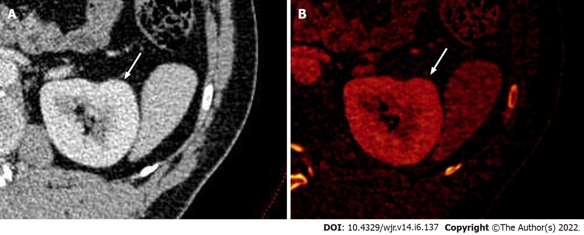
Figure 3 Dual-energy computed tomography images.
A: Contrast-enhanced axial dual-energy computed tomography image showing contour bulge from the lateral cortex of the interpolar region of the left kidney, enhanced similar to background parenchyma; B: Iodine overlay image confirming the absence of any differential iodine distribution, suggesting the lesion to be a dromedary hump.
- Citation: Aggarwal A, Das CJ, Sharma S. Recent advances in imaging techniques of renal masses. World J Radiol 2022; 14(6): 137-150
- URL: https://www.wjgnet.com/1949-8470/full/v14/i6/137.htm
- DOI: https://dx.doi.org/10.4329/wjr.v14.i6.137


