Copyright
©The Author(s) 2016.
World J Radiol. Jun 28, 2016; 8(6): 628-634
Published online Jun 28, 2016. doi: 10.4329/wjr.v8.i6.628
Published online Jun 28, 2016. doi: 10.4329/wjr.v8.i6.628
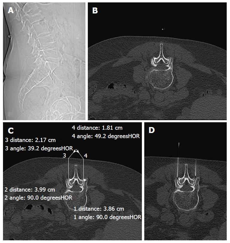
Figure 1 Set of four standard images presented to the experimental group.
A: Sagittal scout view of the lumbar spine; B: Axial CT image showing the degenerated facet joints; C: Axial CT image showing the planned course of the needles; D: CT image showing the final positions of the needle tip on each side. CT: Computed tomography.
- Citation: Middendorp M, Kollias K, Ackermann H, Splettstößer A, Vogl TJ, Khan MF, Maataoui A. Does therapist’s attitude affect clinical outcome of lumbar facet joint injections? World J Radiol 2016; 8(6): 628-634
- URL: https://www.wjgnet.com/1949-8470/full/v8/i6/628.htm
- DOI: https://dx.doi.org/10.4329/wjr.v8.i6.628


