Copyright
©The Author(s) 2016.
World J Radiol. May 28, 2016; 8(5): 449-459
Published online May 28, 2016. doi: 10.4329/wjr.v8.i5.449
Published online May 28, 2016. doi: 10.4329/wjr.v8.i5.449
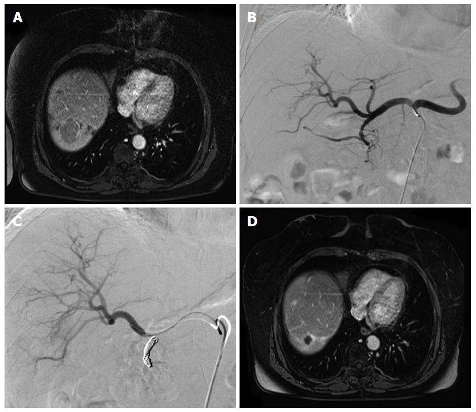
Figure 1 Case study of a 64-year-old female patient suffering from liver metastases from a midgut neuroendocrine tumor.
A: Contrast enhanced MRI shows a large liver metastasis in the right hemiliver; B: Prior to TARE an angiogram of the hepatic arteries was obtained; C: The gastroduodenal artery was occluded with multiple microcoils; D: Contrast enhanced MRI obtained 24 mo after therapy shows a maintained partial response of the liver metastasis. MRI: Magnetic resonance imaging; TARE: Transarterial radioembolization.
- Citation: Mahnken AH. Current status of transarterial radioembolization. World J Radiol 2016; 8(5): 449-459
- URL: https://www.wjgnet.com/1949-8470/full/v8/i5/449.htm
- DOI: https://dx.doi.org/10.4329/wjr.v8.i5.449


