Copyright
©The Author(s) 2016.
World J Radiol. Apr 28, 2016; 8(4): 370-377
Published online Apr 28, 2016. doi: 10.4329/wjr.v8.i4.370
Published online Apr 28, 2016. doi: 10.4329/wjr.v8.i4.370
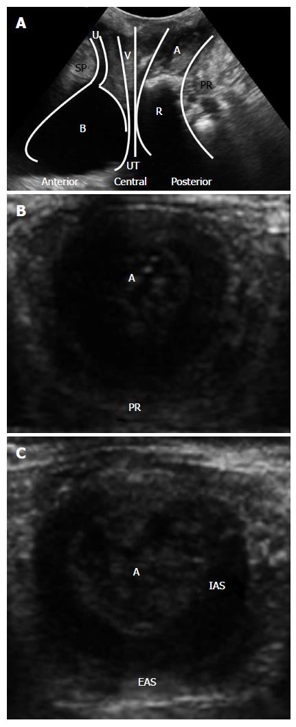
Figure 1 Standard transperineal ultrasound images.
A: Midsagittal view; B: Upper anal canal in the transversal view; C: Transversal view of the middle anal canal. SP: Symphysis pubis; U: Urethra; B: Bladder; V: Vagina; UT: Uterus; R: Rectum; A: Anal canal; PR: Puborectalis muscle; IAS: Internal anal sphincter; EAS: External anal sphincter.
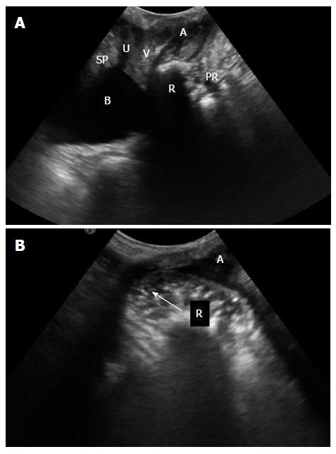
Figure 2 A woman with symptoms of obstructed defecation.
A: Transperineal ultrasound at rest; B: Transperineal ultrasound during maximal straining, after rectum filling with ultrasonographic coupling gel, showing a herniation of the anterior rectal wall into the vagina confirming a retocele (arrow). SP: Symphysis pubis; U: Urethra; B: Bladder; V: Vagina; R: Rectum; A: Anal canal; PR: Puborectalis muscle.
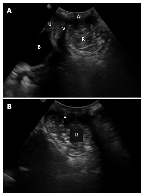
Figure 3 A woman with symptoms of obstructed defecation.
A: Transperineal ultrasound at rest, after rectum filling with ultrasonographic coupling gel; B: Transperineal ultrasound during maximal straining, after rectum filling with ultrasonographic coupling gel, showing a herniation of the rectal wall into the anal canal confirming a rectal intussusception (arrow). U: Urethra; B: Bladder; V: Vagina; R: Rectum; A: Anal canal.
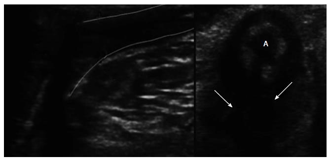
Figure 4 Transversal view of the anal canal (A) in a case of inflammatory perianal disease, fistula (left image, between lines) and abscess (right image, arrow).
- Citation: Albuquerque A, Pereira E. Current applications of transperineal ultrasound in gastroenterology. World J Radiol 2016; 8(4): 370-377
- URL: https://www.wjgnet.com/1949-8470/full/v8/i4/370.htm
- DOI: https://dx.doi.org/10.4329/wjr.v8.i4.370


