Copyright
©The Author(s) 2016.
World J Radiol. Feb 28, 2016; 8(2): 124-131
Published online Feb 28, 2016. doi: 10.4329/wjr.v8.i2.124
Published online Feb 28, 2016. doi: 10.4329/wjr.v8.i2.124
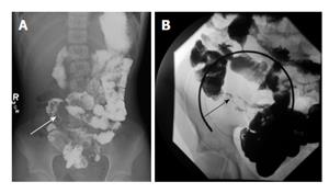
Figure 1 Small bowel follow-through examination in two patients with Crohn’s disease.
A and B demonstrate mucosal irregularity and luminal narrowing of the terminal ileum (arrows).
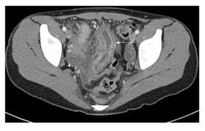
Figure 2 Axial computed tomography image of the pelvis in an adolescent boy with Crohn’s disease.
It demonstrates bowel wall thickening of the distal small bowel and enhancement of the mucosa (arrow). There is surrounding free fluid (arrowhead).
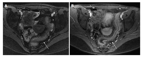
Figure 3 Magnetic resonance enterography study of the pelvis in an adolescent patient with active Crohn’s disease.
T2 weighted image (A) demonstrating free fluid (arrowhead) and bowel wall thickening (arrow); T1 weighted image (B) with contrast demonstrating enhancement of the bowel and increased mesenteric vascularity (arrow).
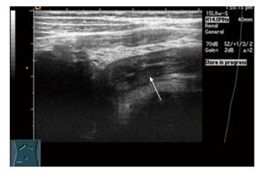
Figure 4 Transabdominal ultrasound in a 12-year-old boy with Crohn’s disease.
It demonstrates bowel wall thickening of the terminal ileum.
- Citation: Haas K, Rubesova E, Bass D. Role of imaging in the evaluation of inflammatory bowel disease: How much is too much? World J Radiol 2016; 8(2): 124-131
- URL: https://www.wjgnet.com/1949-8470/full/v8/i2/124.htm
- DOI: https://dx.doi.org/10.4329/wjr.v8.i2.124


