Copyright
©The Author(s) 2016.
World J Radiol. Oct 28, 2016; 8(10): 829-845
Published online Oct 28, 2016. doi: 10.4329/wjr.v8.i10.829
Published online Oct 28, 2016. doi: 10.4329/wjr.v8.i10.829
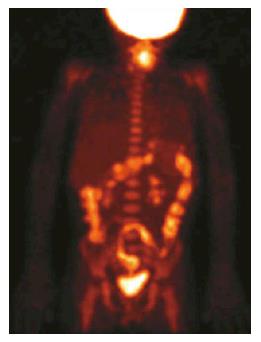
Figure 1 Maximum intensity projection image of an positron emission tomography with 18F-Fluorodehoxiglucose examination shows diffuse and intense radiopharmaceutical uptake in the large bowel.
Reprinted with permission of Springer Verlag from Cistaro et al.
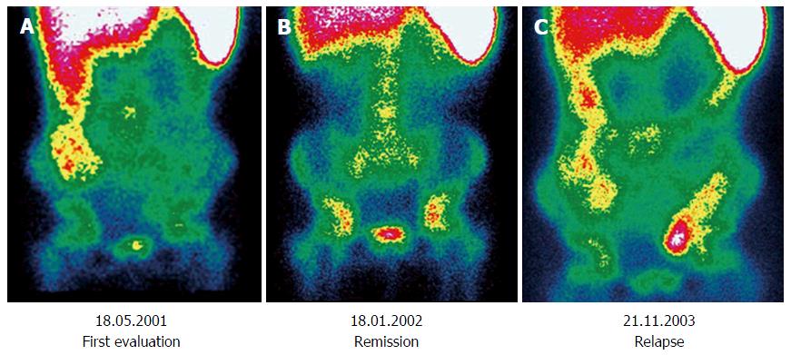
Figure 2 Three sequential 99mTc-HMPAO-white blood cell scintigraphies performed in a patient diagnosed with Crohn’s disease.
A: Performed at diagnosis, there is evidence of increased activity involving colon ascendens; B: Performed at follow-up after immunological therapy, a complete remission can be demonstrated; C: There is evidence of a relapse involving colon ascendens and sigma-rectum. Reprinted with permission of Springer Verlag from Caobelli et al[34].
- Citation: Caobelli F, Evangelista L, Quartuccio N, Familiari D, Altini C, Castello A, Cucinotta M, Di Dato R, Ferrari C, Kokomani A, Laghai I, Laudicella R, Migliari S, Orsini F, Pignata SA, Popescu C, Puta E, Ricci M, Seghezzi S, Sindoni A, Sollini M, Sturiale L, Svyridenka A, Vergura V, Alongi P, Young AIMN Working Group. Role of molecular imaging in the management of patients affected by inflammatory bowel disease: State-of-the-art. World J Radiol 2016; 8(10): 829-845
- URL: https://www.wjgnet.com/1949-8470/full/v8/i10/829.htm
- DOI: https://dx.doi.org/10.4329/wjr.v8.i10.829


