Copyright
©The Author(s) 2015.
World J Radiol. Dec 28, 2015; 7(12): 501-508
Published online Dec 28, 2015. doi: 10.4329/wjr.v7.i12.501
Published online Dec 28, 2015. doi: 10.4329/wjr.v7.i12.501
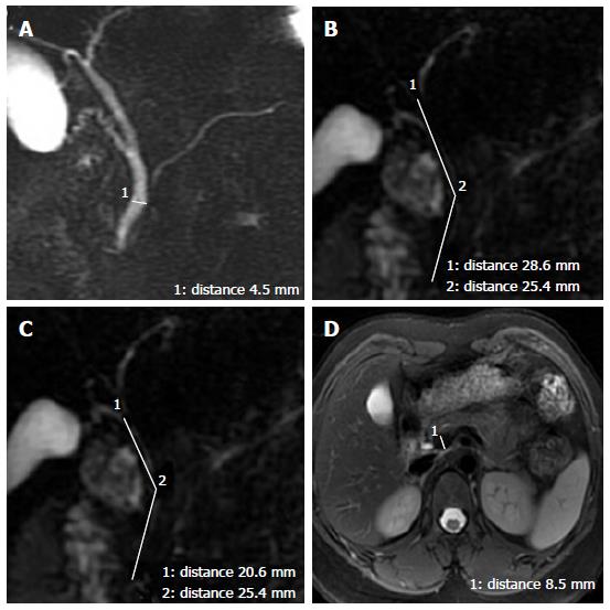
Figure 1 The measurement method.
A: Measurement of the common bile duct (CBD) diameter by placing an electronic caliper at the widest visible portion of the CBD on magnetic resonance cholangiopancreatography (MRCP); B: Measurement of the length of the extrahepatic bile duct on MRCP. It is the sum of the length from the hepatic hilum to the tortuous portion and from the tortuous portion to the ampulla; C: Measurement of the length of the CBD on MRCP. It is the sum of the length from the cystic duct insertion to the tortuous portion and from the tortuous portion to the ampulla; D: Measurement of the portal vein anteroposterior diameters by placing the electronic caliper at the splenic veins into the portal vein on T2-weighted images.
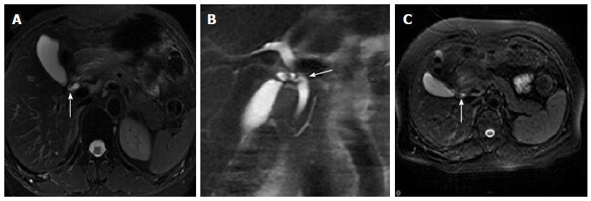
Figure 2 The cystic junction radial orientation.
An FRFSE T2-weighted image (A) shows lateral insertion of the cystic duct (arrow). A coronal SSFSE T2-weighted image (B) shows medial insertion of the cystic duct (arrow). An FRFSE T2-weighted image (C) shows posteroanterior insertion of the cystic duct (arrow). SSFSE: Single-shot fast spin-echo; FRFSE: Fast-recovery fast-spin echo.
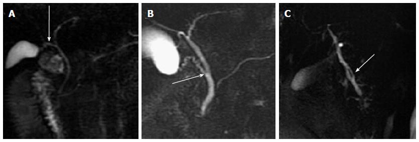
Figure 3 The cystic junction location.
Magnetic resonance cholangiopancreatography shows proximal (A), middle (B) and distal (C) third conjunction of the cystic duct with the common bile duct (arrow).
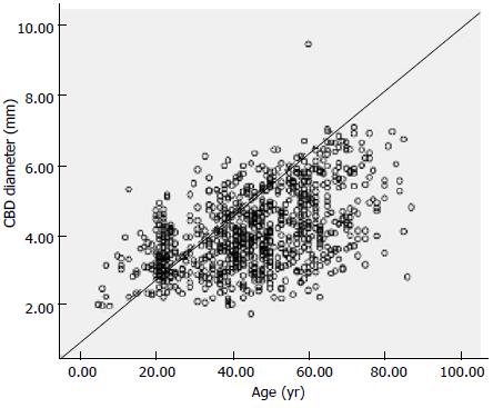
Figure 4 Pearson correlation between the diameter of the common bile duct and age (r = 0.
484, P = 0.000).
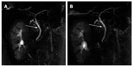
Figure 5 Deep respiratory magnetic resonance cholangiopancreatography obtained in a 32-year-old female volunteer.
Breath-hold magnetic resonance cholangiopancreatography obtained during end-expiration (A) or end-inspiration (B) provides an overview of the common bile duct (arrow). There is no obvious change in the common bile duct diameter.
- Citation: Peng R, Zhang L, Zhang XM, Chen TW, Yang L, Huang XH, Zhang ZM. Common bile duct diameter in an asymptomatic population: A magnetic resonance imaging study. World J Radiol 2015; 7(12): 501-508
- URL: https://www.wjgnet.com/1949-8470/full/v7/i12/501.htm
- DOI: https://dx.doi.org/10.4329/wjr.v7.i12.501


