Copyright
©2014 Baishideng Publishing Group Co.
World J Radiol. Apr 28, 2014; 6(4): 119-124
Published online Apr 28, 2014. doi: 10.4329/wjr.v6.i4.119
Published online Apr 28, 2014. doi: 10.4329/wjr.v6.i4.119
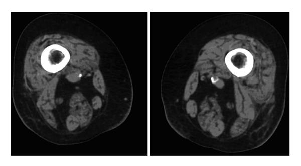
Figure 1 Cross-sectional computed tomography at the level of the middle thigh showing fatty degeneration and atrophy of skeletal muscles in a vitamin D-deficient patient.
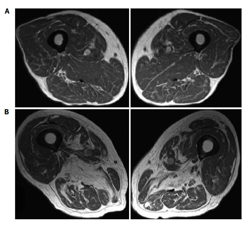
Figure 2 Magnetic resonance imaging of two different grades of fatty degeneration and atrophy involving thigh muscle in patients with vitamin D deficiency.
A: Grade 1 (less than 30% of the volume of muscles involved); B: Grade 2 (30%-60% of the volume of muscles compromised).
- Citation: Bignotti B, Cadoni A, Martinoli C, Tagliafico A. Imaging of skeletal muscle in vitamin D deficiency. World J Radiol 2014; 6(4): 119-124
- URL: https://www.wjgnet.com/1949-8470/full/v6/i4/119.htm
- DOI: https://dx.doi.org/10.4329/wjr.v6.i4.119


