Copyright
©2014 Baishideng Publishing Group Co.
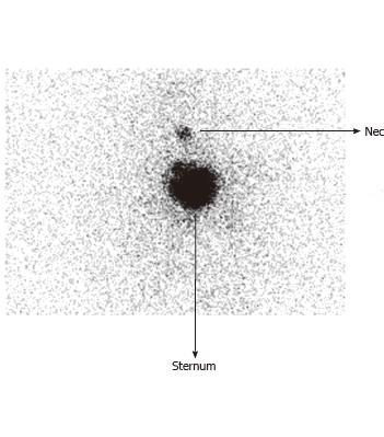
Figure 1 Pre-treatment diagnostic radioiodine scan (post thyroidectomy at 5 wk) demonstrating neck uptake and iodine avid sternal lesion.
No other abnormal focus was noted elsewhere in the body.
Figure 2 Post therapy scan while discharging the patient from radioiodine therapy ward demonstrates a left abdominal focal lesion in addition to the neck and sternal focus.
Figure 3 Post-therapy scan demonstrating the left sided abdominal focus more vividly.
A: Planar spot view of abdomen; B: Single-photon emission computed tomography (SPECT) coronals (SPECT images); C: The 3-slice SPECT display (transaxials, sagittals, coronals): Arrow indicates the adrenal metastasis.
Figure 4 Positron emission tomography-computed tomography fusion images demonstrating avid fludeoxyglucose uptake in the left adrenal lesion.
A: Transaxial fludeoxyglucose-PET/CT (CT, PET, fused PET-CT); B: Fused Coronals. PET/CT: Positron emission tomography/computed tomography.
- Citation: Ranade R, Thapa P, Basu S. Adrenal metastasis from differentiated thyroid carcinoma documented on post-therapy 131I scan: A case based discussion. World J Radiol 2014; 6(3): 56-61
- URL: https://www.wjgnet.com/1949-8470/full/v6/i3/56.htm
- DOI: https://dx.doi.org/10.4329/wjr.v6.i3.56


