Copyright
©2014 Baishideng Publishing Group Inc.
World J Radiol. Oct 28, 2014; 6(10): 794-807
Published online Oct 28, 2014. doi: 10.4329/wjr.v6.i10.794
Published online Oct 28, 2014. doi: 10.4329/wjr.v6.i10.794
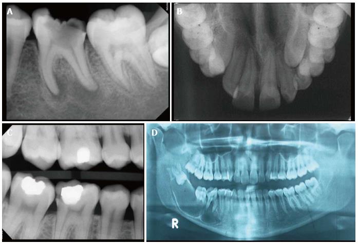
Figure 1 X-ray.
A: An intraoral periapical X-ray helps to view the number and morphology of roots and root canals, periapical status and alveolar bone support interdentally. In this case, a grossly carious lower molar with diffuse radiolucency around both the root apices, denoting a chronic abscess is seen; B: An occlusal view of maxilla is useful to evaluate suture line of maxillary processes and extent of large pathological lesion such as a cyst and for location of impacted teeth. In this X-ray an impacted left maxillary canine can be seen; C: A bitewing X-ray is useful to examine a segment of upper and lower arch simultaneously to detect inter-proximal caries. In this X-ray, distal proximal surface of the lower 1st molar shows a carious defect; D: A panoramic radiograph gives a bird‘s-eye view of upper and lower jaws with excellent view of temporo-mandibular joint and maxillary sinuses. In this X-ray, a large, multi-locular cystic lesion, involving an impacted and inverted 3rd molar, extending up to the premolar region can be seen.
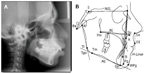
Figure 2 Cephalometric radiographs show the entire side of the head and help to evaluate the spatial relationships between cranial and dental structures.
A: A lateral cephalometric X-ray is useful to determine cranio-facial structures and their relationship with position of the jaws and teeth; B: Different landmarks used to evaluate the planes and angles formed, to arrive at a diagnosis and treatment planning for orthodontic treatment/orthognathic surgery. Serial cephalograms can give the amount and direction of growth of facio-maxillary complex.
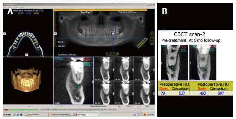
Figure 3 Cone beam computed tomography.
A: A cone beam computed tomography scan gives a three-dimensional view of the area of interest. In this case, the periapical lesion is being evaluated; B: The image gives values in Hounsfield unit of cementum and alveolar bone density to measure post-treatment healing. CBCT: Cone beam computed tomography.
- Citation: Shah N, Bansal N, Logani A. Recent advances in imaging technologies in dentistry. World J Radiol 2014; 6(10): 794-807
- URL: https://www.wjgnet.com/1949-8470/full/v6/i10/794.htm
- DOI: https://dx.doi.org/10.4329/wjr.v6.i10.794


