Copyright
©2013 Baishideng Publishing Group Co.
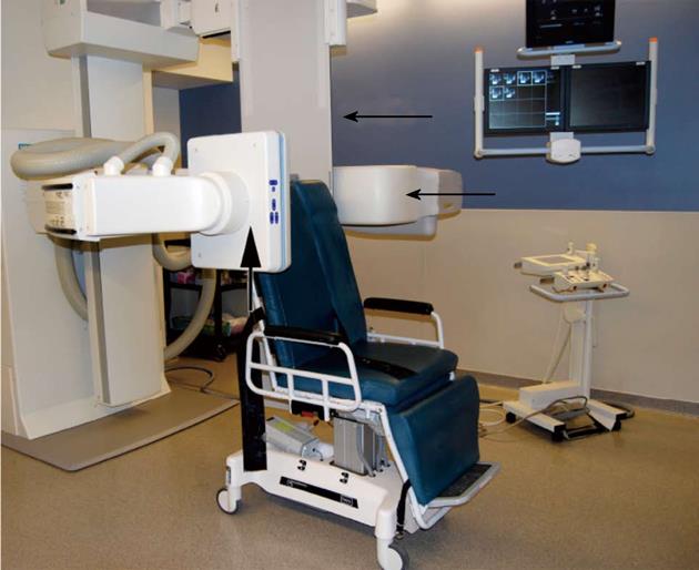
Figure 1 Siemens artis fluoroscopy suite.
The table is set up for an upper gastrointestinal (GI) study. Note the relationship of the image intensifier to the table (arrows). The most likely cause of the infections was contamination of the image intensifier and fluoroscopy table during these studies with inadequate cleaning of the suite between GI procedures and arthrograms.
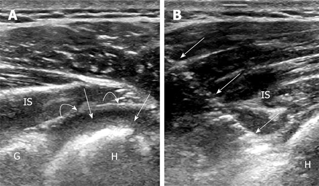
Figure 2 Ultrasound image through the posterior shoulder.
A: Axial ultrasound image through the posterior shoulder shows a large joint effusion (straight arrows) with fluid between the humeral head (H) and the adjacent posterior capsule (curved arrows); B: Ultrasound guided aspiration of the posterior shoulder. Note the needle (arrows). Cultures grew Streptococcus crista. IS: infraspinatus; G: Posterior glenoid.
- Citation: Vollman AT, Craig JG, Hulen R, Ahmed A, Zervos MJ, Holsbeeck MV. Review of three magnetic resonance arthrography related infections. World J Radiol 2013; 5(2): 41-44
- URL: https://www.wjgnet.com/1949-8470/full/v5/i2/41.htm
- DOI: https://dx.doi.org/10.4329/wjr.v5.i2.41


