Copyright
©2012 Baishideng Publishing Group Co.
World J Radiol. Aug 28, 2012; 4(8): 387-390
Published online Aug 28, 2012. doi: 10.4329/wjr.v4.i8.387
Published online Aug 28, 2012. doi: 10.4329/wjr.v4.i8.387
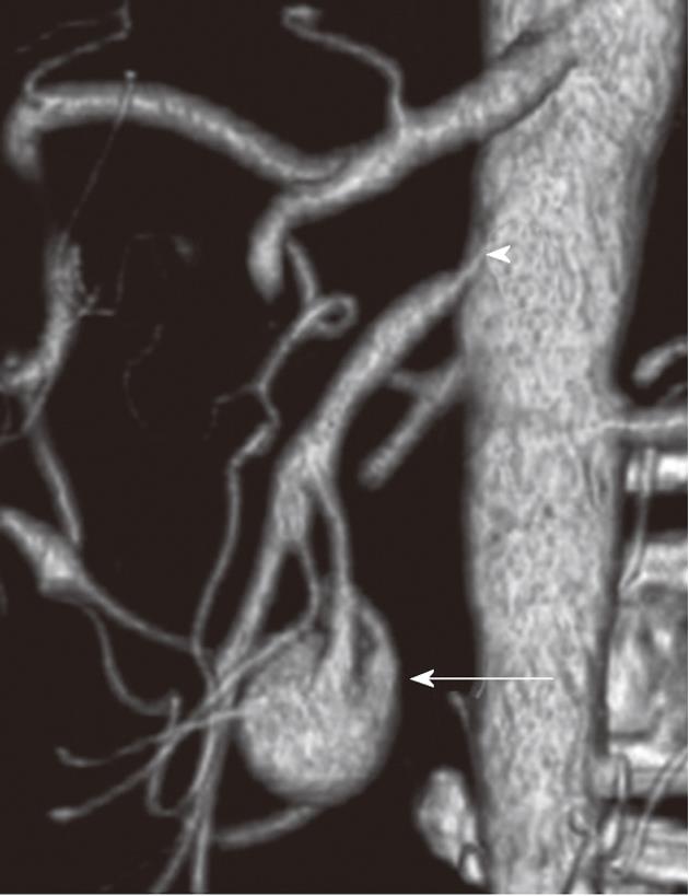
Figure 1 Volume-rendered three-dimensional computed tomography demonstrates an aneurysm (arrow) supplied from inferior pancreaticoduodenal artery and the overt stenosis of superior mesenteric artery at origin (arrowhead).
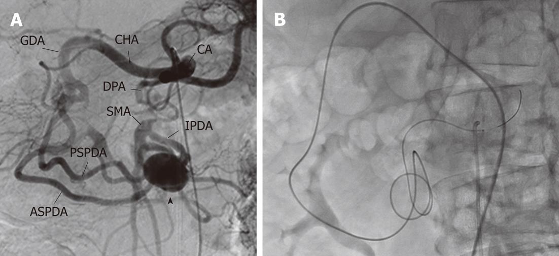
Figure 2 Angiography and catheterization prior to treatment.
A: Celiac arteriography demonstrates a superior mesenteric artery via the dilated posterior and anterior pancreaticoduodenal arcades with aneurysm (black arrow head); B: Successful catheterization was achieved using a microwire, a microcatheter and Roesch celiac (RC) catheter from celiac artery to superior mesenteric artery and abdominal aorta via anterior pancreaticoduodenal arcade and an aneurysm. CA: Celiac artery; CHA: Common hepatic artery; GDA: Gastroduodenal artery; DPA: Dorsal pancreatic artery; SMA: Superior mesenteric artery; IPDA: Inferior pancreaticoduodenal artery; PSPDA: Posterior superior pancreaticoduodenal artery; ASPDA: Anterior superior pancreaticoduodenal artery.
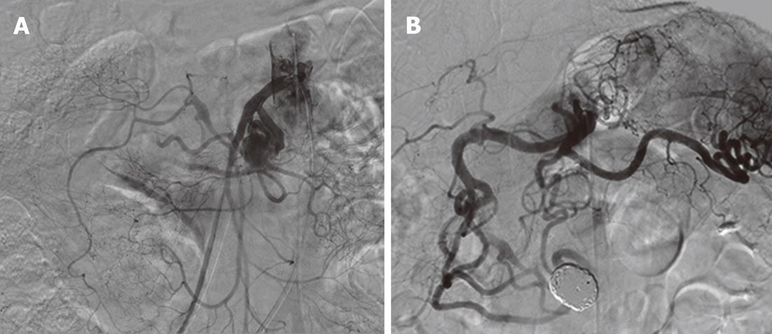
Figure 3 Angiography following stent placement and microcoils embolization.
A: Superior mesenteric arteriography following stent placement demonstrates the dilated superior mesenteric artery at origin; B: Celiac arteriography following microcoils embolization via superior mesenteric artery demonstrates the eradication of the aneurysm. An arrow indicates a metallic stent placed at the origin of the superior mesenteric artery.
- Citation: Ikoma A, Nakai M, Sato M, Kawai N, Tanaka T, Sanda H, Nakata K, Minamiguchi H, Sonomura T. Inferior pancreaticoduodenal artery aneurysm treated with coil packing and stent placement. World J Radiol 2012; 4(8): 387-390
- URL: https://www.wjgnet.com/1949-8470/full/v4/i8/387.htm
- DOI: https://dx.doi.org/10.4329/wjr.v4.i8.387


