Copyright
©2012 Baishideng Publishing Group Co.
World J Radiol. Jul 28, 2012; 4(7): 302-310
Published online Jul 28, 2012. doi: 10.4329/wjr.v4.i7.302
Published online Jul 28, 2012. doi: 10.4329/wjr.v4.i7.302
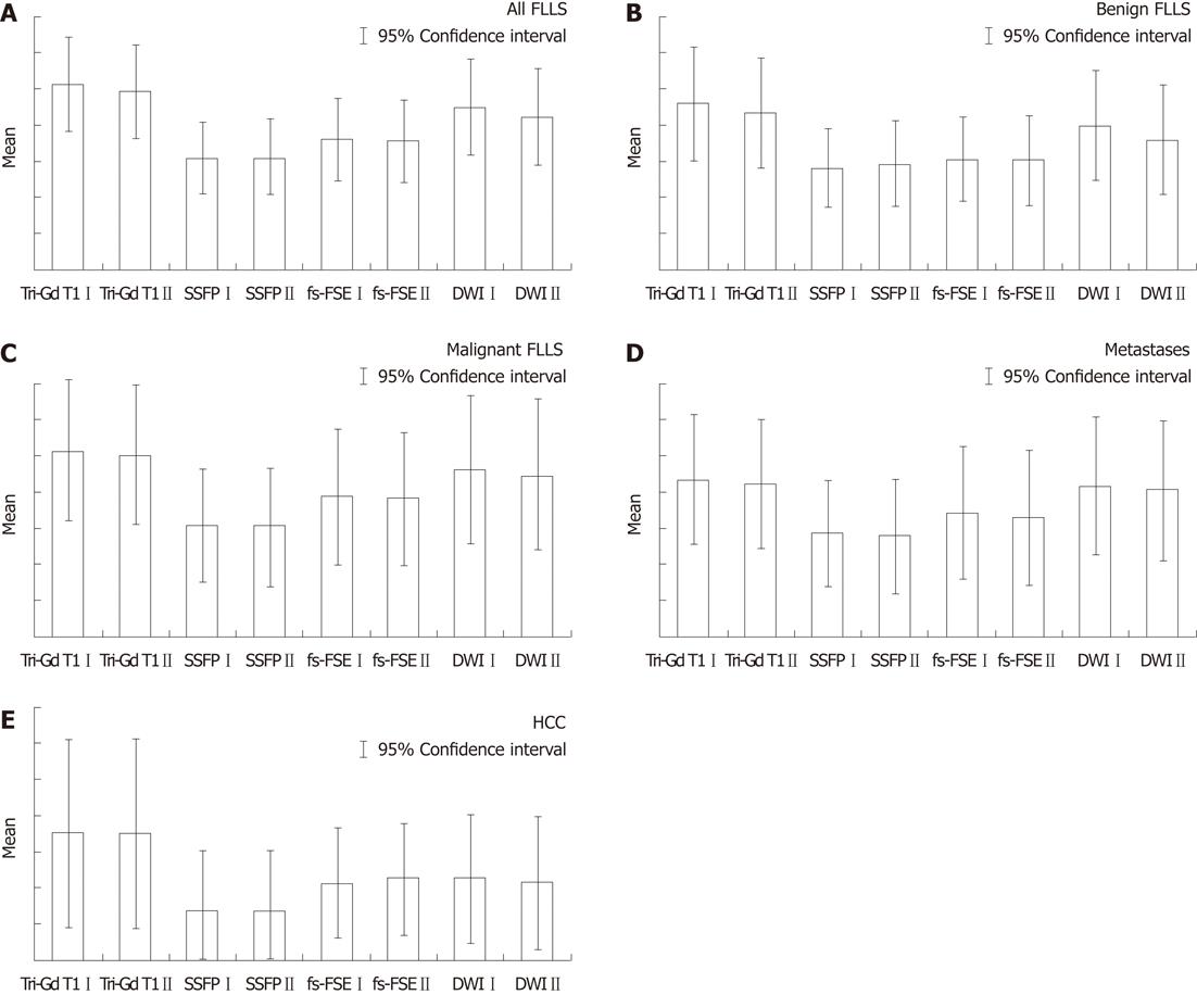
Figure 1 Histograms comparing the different sequences (A-E).
Histograms showing the comparison between the mean values of lesions detected among different sequences, for the classes of focal liver lesions (FLLs) [all lesions, benign lesions, malignant lesions, metastases, hepatocellular carcinomas (HCCs)]. I= reader 1; II = reader 2; Tri-Gd T1: Triphasic gadolinium T1-weighted sequence; fs-FSE: Fat suppressed fast spin-echo sequence; SSFP: Steady state free precession; DWI: Diffusion weighted imaging.
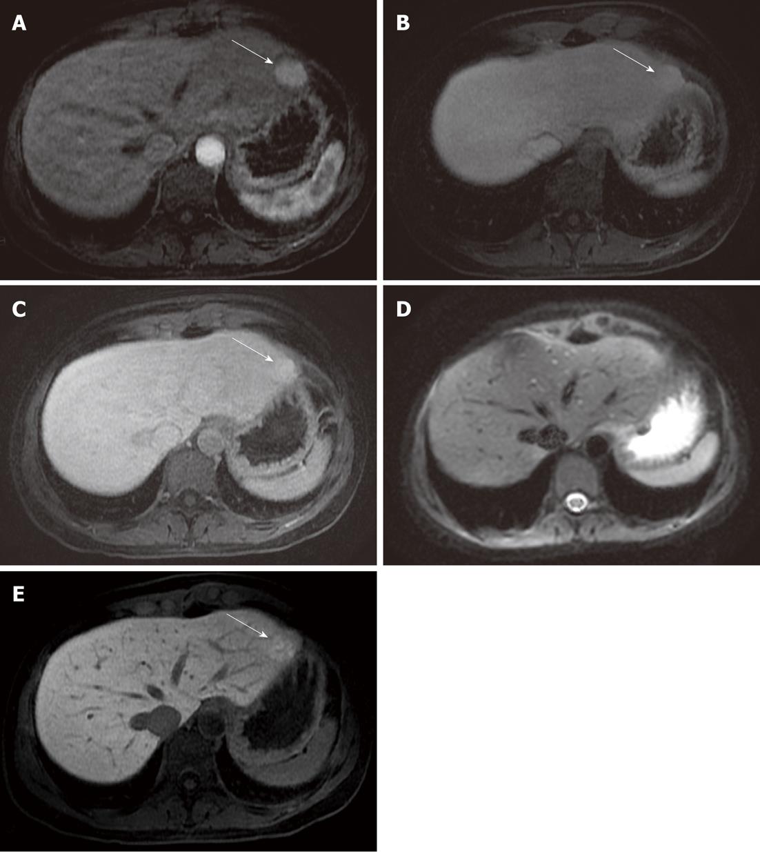
Figure 2 Focal nodular hyperplasia found in the left liver lobe (arrow).
A: A round-shaped solid lesion (arrow) is depicted in the left liver lobe (in the II segment); B, C: The lesion (arrow) appears homogeneously hyperintense in the arterial phase, and remains slightly hyperintense in the portal and in the equilibrium phase; D: The lesion was missed on the diffusion image by both readers, probably due to its location near the gastric wall, along the liver surface; E: In the hepato-specific phase, the lesion (arrow) shows uptake of gadolinium-ethoxybenzyl-diethylenetriamine pentaacetic acid; a diagnosis of focal nodular hyperplasia was suggested due to the dynamic behavior observed after contrast administration.
Figure 3 Hepatocellular carcinoma in the left liver lobe.
A: Hypervascular lesions depicted (arrows) in the arterial phase; B: Lesions show wash-out in the venous phase (arrows), suggesting the diagnosis of hepatocellular carcinomas; C: On diffusion imaging, acquired at b10 value, lesions were not detected by the readers, due to their location near the gastric wall.
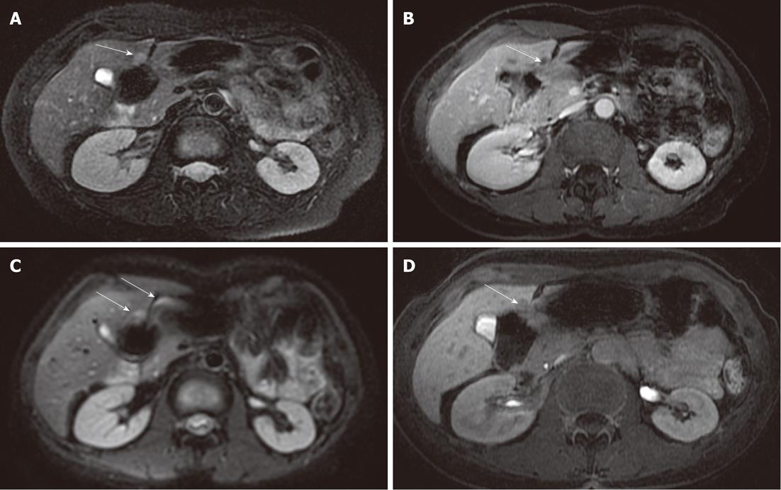
Figure 4 Metastasis in the IV segment.
A: A metastatic lesion in colon carcinoma is depicted in the most caudal area of the IV liver segment, it was well depicted in the fat-suppressed fast spin-echo T2-weighted sequence (arrow); B: In the gadolinium-enhanced acquisition (arrow); C: The lesion is poorly represented in the diffusion-weighted imaging, covered by magnetic susceptibility artifacts due to the adjacent intestinal loop (arrows); D: Delayed imaging after gadolinium-ethoxybenzyl-diethylenetriamine pentaacetic acid confirmed the metastatic lesion (arrow).
- Citation: Palmucci S, Mauro LA, Messina M, Russo B, Failla G, Milone P, Berretta M, Ettorre GC. Diffusion-weighted MRI in a liver protocol: Its role in focal lesion detection. World J Radiol 2012; 4(7): 302-310
- URL: https://www.wjgnet.com/1949-8470/full/v4/i7/302.htm
- DOI: https://dx.doi.org/10.4329/wjr.v4.i7.302


