Copyright
©2012 Baishideng Publishing Group Co.
World J Radiol. Jun 28, 2012; 4(6): 286-290
Published online Jun 28, 2012. doi: 10.4329/wjr.v4.i6.286
Published online Jun 28, 2012. doi: 10.4329/wjr.v4.i6.286
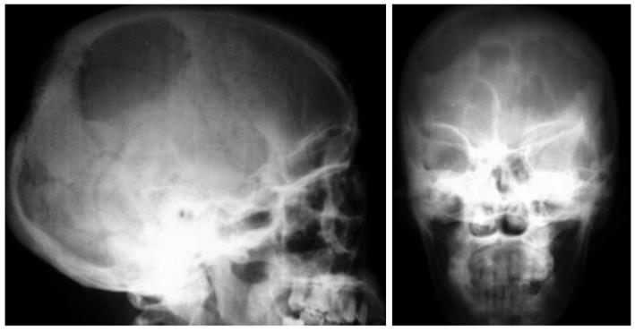
Figure 1 X-ray of skull showing a couple of painless, progressively increasing swellings in the occipitoparietal region of the scalp.
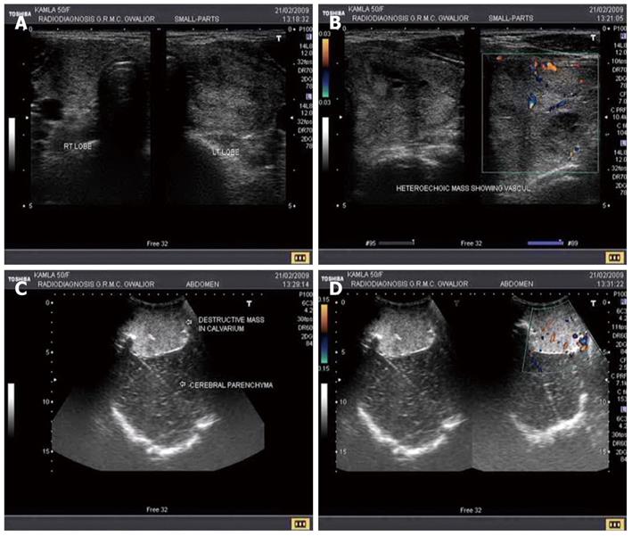
Figure 2 Ultrasound image.
A, B: The ultrasound image of palpable swelling in the neck revealing a heteroechoic lesion; C, D: The ultrasound image of scalp showing a destructive mass in the skull with increased vascularity.
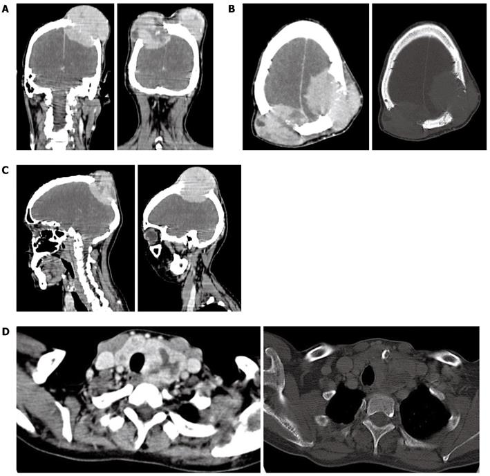
Figure 3 Computed tomography image.
A-C: Computed tomography (CT) image of head revealing defect in the calvarium with a soft tissue density lesion having both intra- as well as extracranial soft tissue components; D: CT image of neck shows large mass involving whole of neck, trachea and vessels.
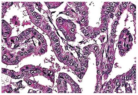
Figure 4 Histopathological report of biopsy from the thyroid lesion revealing branching papillae having a dense fibrovascular core covered with cuboidal epithelial cells that have nuclei with a clear ground glass appearance.
- Citation: Nigam A, Singh AK, Singh SK, Singh N. Skull metastasis in papillary carcinoma of thyroid: A case report. World J Radiol 2012; 4(6): 286-290
- URL: https://www.wjgnet.com/1949-8470/full/v4/i6/286.htm
- DOI: https://dx.doi.org/10.4329/wjr.v4.i6.286


