Copyright
©2012 Baishideng Publishing Group Co.
World J Radiol. May 28, 2012; 4(5): 228-230
Published online May 28, 2012. doi: 10.4329/wjr.v4.i5.228
Published online May 28, 2012. doi: 10.4329/wjr.v4.i5.228
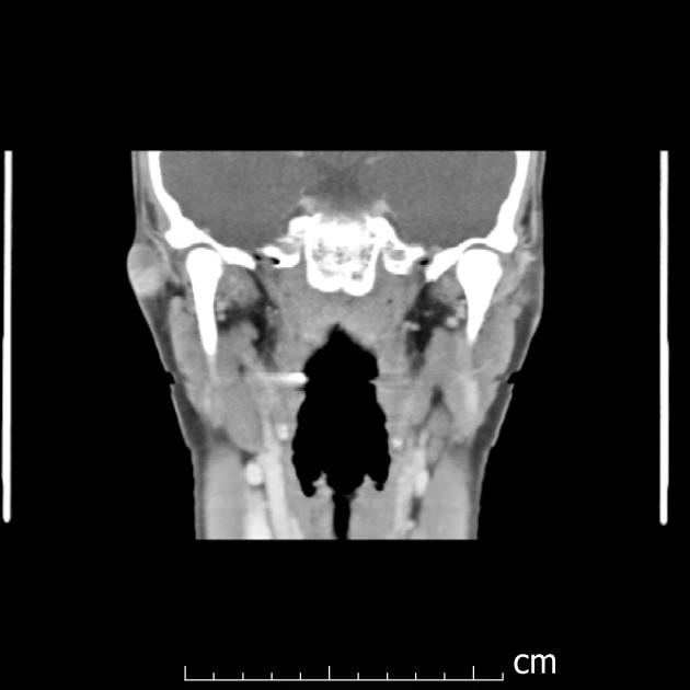
Figure 1 Coronal computed tomographic scan with contrast of the facial region.
This demonstrates a lesion in the pre-auricular region.
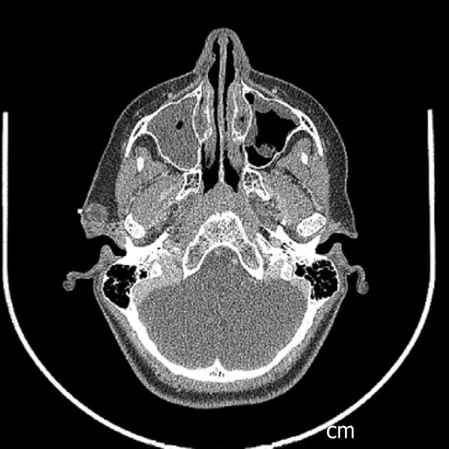
Figure 2 Axial computed tomographic scan with contrast of the facial region.
Soft tissue windows demonstrate the lesion in the pre-auricular region.
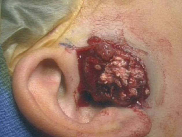
Figure 3 Interoperative photograph of vascular lesion with calcification, containing cheesy inclusions.
It was determined to be a pilomatrixoma.
- Citation: Whittemore KR, Cohen M. Imaging and review of a large pre-auricular pilomatrixoma in a child. World J Radiol 2012; 4(5): 228-230
- URL: https://www.wjgnet.com/1949-8470/full/v4/i5/228.htm
- DOI: https://dx.doi.org/10.4329/wjr.v4.i5.228


