Copyright
©2012 Baishideng Publishing Group Co.
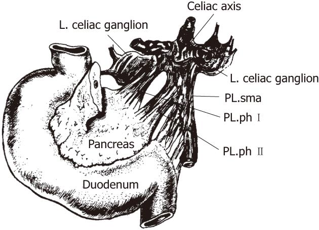
Figure 1 The anatomy of the extrapancreatic plexus[4].
PL: Plexus; sma: Superior mesenteric artery; ph: Pancreatic head.
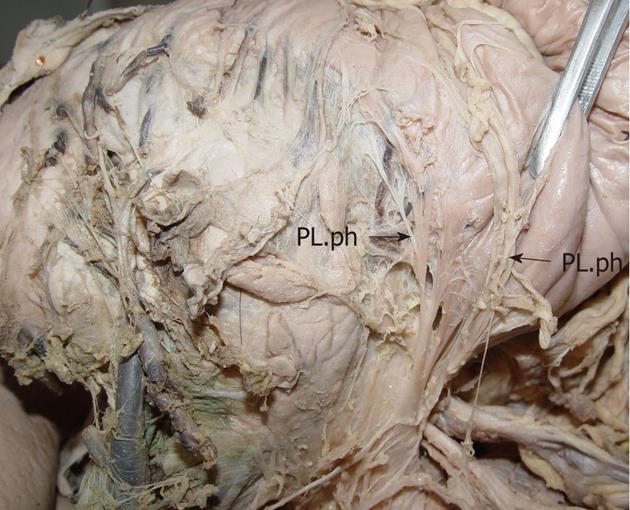
Figure 2 The anatomy of the pancreatic head plexus in a cadaver (arrows).
The plexus extends from the celiac ganglia to the posterior surface of the head of the pancreas, the uncinate process. PL.ph: The plexus of the pancreatic head.
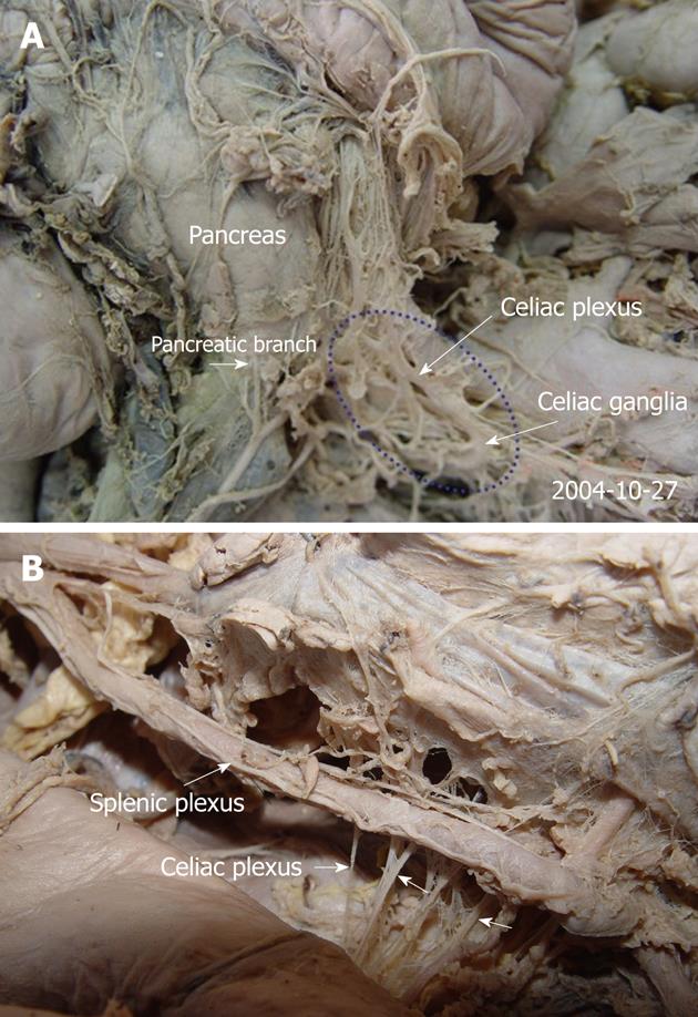
Figure 3 The anatomy of the celiac plexus in cadavers (A, B).
The pancreas was moved upward. The pictures demonstrate that the celiac plexus is the center of the viscus and composed of the celiac ganglia and several large and small nerves that terminate with the celiac ganglia (arrows).
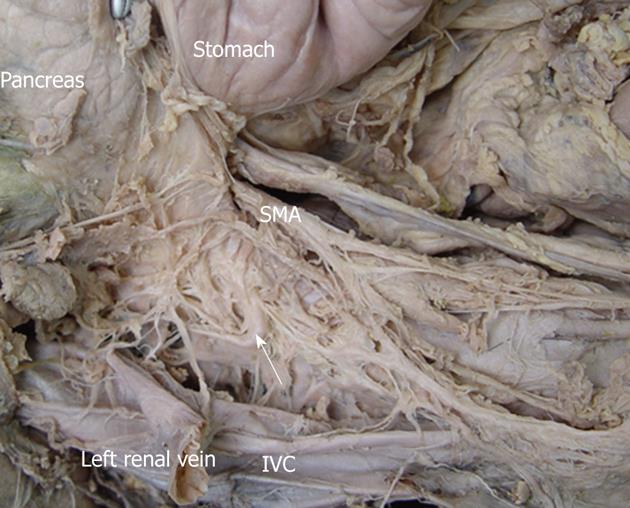
Figure 4 The anatomy of the celiac ganglia in a cadaver.
The celiac ganglion (white arrows) was close to the aorta at a level intermediate to the origin of the celiac artery and superior to the mesenteric artery. It was located in the space bound by the inferior vena cava (IVC), the head of pancreas and the superior mesenteric artery (SMA). The pancreas was moved upward. The IVC was cut and moved laterally.
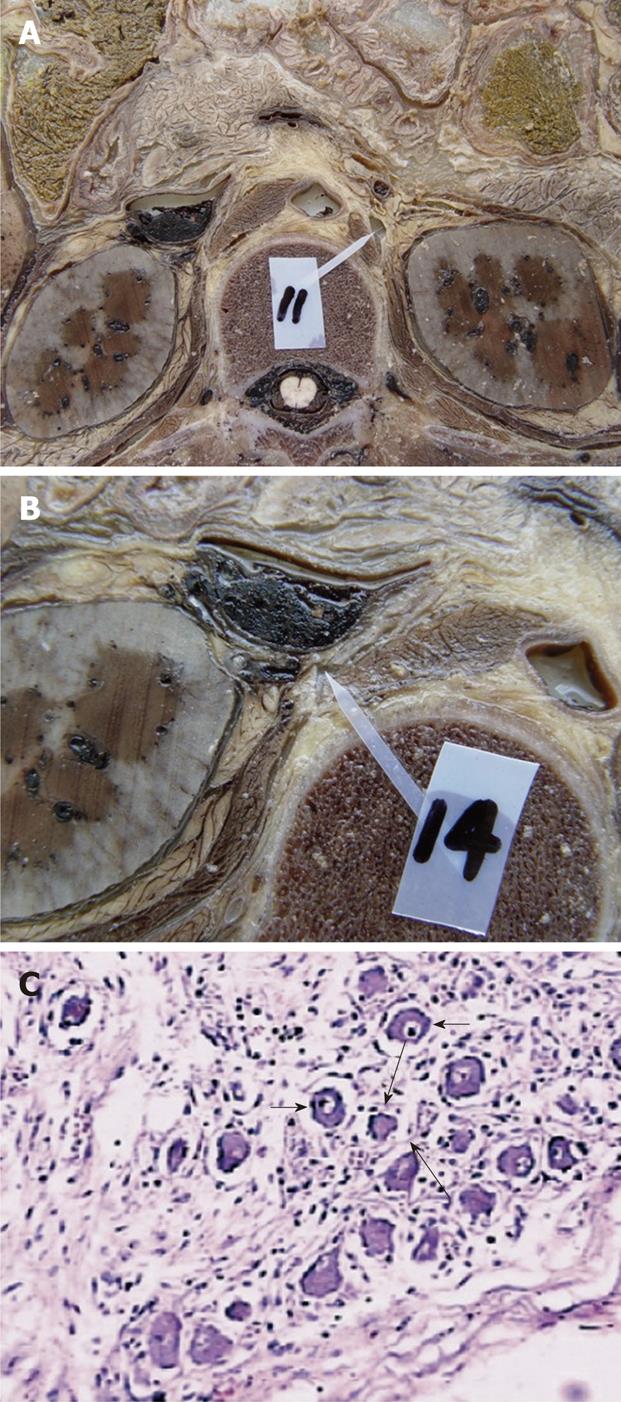
Figure 5 The dissection of the celiac ganglia in a cadaver at the L1 level.
A: The left celiac ganglia; B: The right celiac ganglia; C: The histologic specimen stained with hematoxylin-eosin staining (× 100). With light microscopy, the celiac ganglion shows scattered ganglion cells (short arrows) and sparse nerve fibers (long arrows) among these ganglion cells.
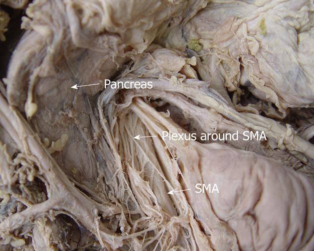
Figure 6 The anatomy of the plexus around the superior mesenteric artery in a cadaver.
This route extends from the superior mesenteric artery (SMA) to the right edge of incisure of the pancreas.
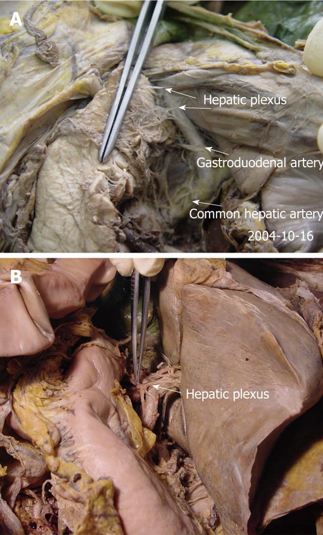
Figure 7 The anatomy of the hepatic plexus in cadavers (A, B).
This route goes from the liver to the back and superior border of the pancreatic head, along the common hepatic and gastroduodenal artery (arrows).
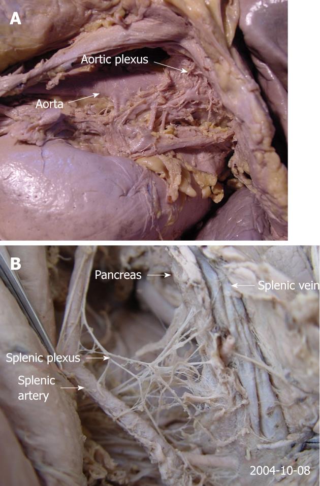
Figure 8 The anatomy of the aortic plexus in a cadaver.
A: This route and its branches surround the aorta and extend to the head and uncinate process of the pancreas; B: The route travels along the splenic artery, and the main route and its many branches reach the tail of the pancreas.
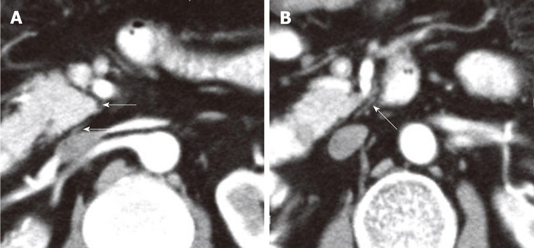
Figure 9 A contrast-enhanced computed tomography shows the plexus (PLX-II) (A, B).
The PLX-II extends to the left margin of the uncinate process via the plexus surrounding the superior mesenteric artery (arrows).
Figure 10 Non-enhanced computed tomography and contrast-enhanced computed tomography images show the neural plexus of the head of the pancreas (A-C).
The plexus was a strand-like structure located in the area bound by the superior mesenteric artery, the inferior vena cava and the abdominal aorta (arrows).
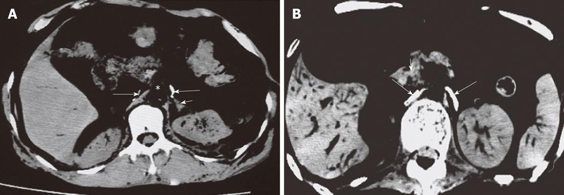
Figure 11 Computed tomography images of celiac ganglion in cadavers (labeled with contras media).
The left was crescent-shaped and in front of the left adrenal gland, and the right was line-shaped and adjacent to the right crura of diaphragm (long arrows). Short arrows indicate left adrenal gland (A) and superior mesenteric artery (B).
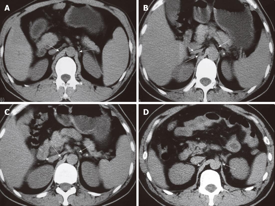
Figure 12 Computed tomography images show the celiac ganglia at different levels.
A: A celiac ganglion (long arrows) at the root of the superior mesenteric artery; its density is the same as the liver and spleen or slightly less than the diaphragma crura; B: The celiac ganglia (long arrows) at the level of the pancreatic head and neck. The left celiac ganglia were lamina-shaped and located lengthwise in the space in the front of the left adrenal gland (*); the right celiac ganglia were a thick, line-shaped structure dorsal to the inferior vena cava (IVC) and lateral to the right diaphragma crura; C: A celiac ganglion (long arrows) at the level of the root of the celiac trunk. It appeared lamina- and nodule-shaped; D: The celiac ganglia (long arrows) at the level of the uncinate process, with well-defined margins. The left ganglia were lamina-shaped and located lengthwise in the space to the front and the left of the adrenal gland (*); the right ganglia were thick and line-shaped, located dorsal to the IVC and lateral to the right diaphragma crura. Short arrows indicate IVC (B, C).
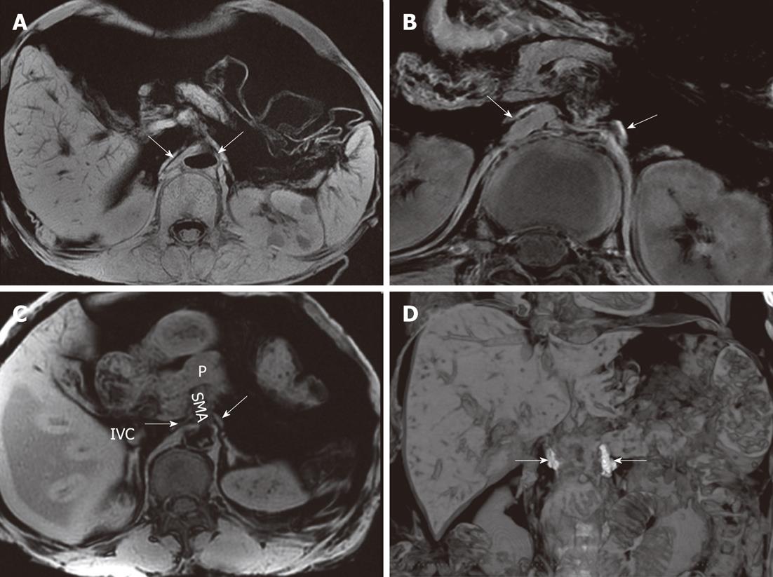
Figure 13 The celiac ganglia on an magnetic resonance imaging.
A: A gradient-refocused-echo T1-weighted image shows that both the right and left celiac ganglia (arrows), labeled with Gd-DTPA, have a higher signal intensity than that of the viscus, such as the liver and spleen; B: 3D T1-weighted image shows that both the right and left celiac ganglia labeled with Gd-DTPA (arrows) have a higher signal intensity than the kidneys; C: A gradient-refocused-echo, T1-weighted, out-of-phase image shows the right and left celiac ganglia (labeled with arrows). The right celiac ganglion was located in the space formed by the inferior vena cava (IVC), right adrenal gland, right diaphragmatic crura, head of pancreas, and superior mesenteric artery (SMA). The left ganglion was located in the open space formed by the left adrenal gland, left diaphragmatic crura, and SMA. The celiac ganglia are labeled with gadolinium; D: Coronal imaging on T1-weighted images shows the celiac ganglia labeled with Gd-DTPA (arrows). P: Pancreas.
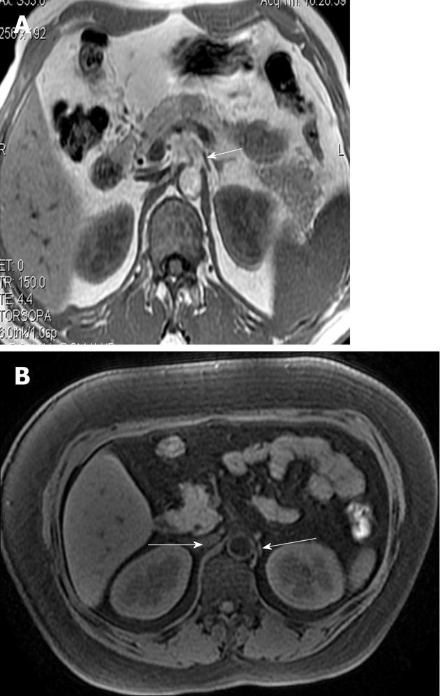
Figure 14 The magnetic resonance imagings of normal adults clearly show the celiac ganglia (A, B).
Both celiac ganglia were seen at the level between the celiac artery and the superior mesenteric artery (arrows).
- Citation: Zuo HD, Zhang XM, Li CJ, Cai CP, Zhao QH, Xie XG, Xiao B, Tang W. CT and MR imaging patterns for pancreatic carcinoma invading the extrapancreatic neural plexus (Part I): Anatomy, imaging of the extrapancreatic nerve. World J Radiol 2012; 4(2): 36-43
- URL: https://www.wjgnet.com/1949-8470/full/v4/i2/36.htm
- DOI: https://dx.doi.org/10.4329/wjr.v4.i2.36


