Copyright
©2011 Baishideng Publishing Group Co.
World J Radiol. Jul 28, 2011; 3(7): 178-181
Published online Jul 28, 2011. doi: 10.4329/wjr.v3.i7.178
Published online Jul 28, 2011. doi: 10.4329/wjr.v3.i7.178
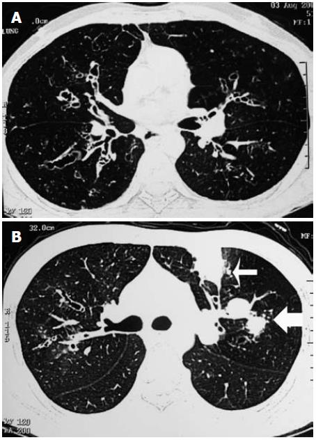
Figure 1 High-resolution computed tomography of the chest from two different patients showing typical central bronchiectasis (panel A).
Mucus plugging within dilated bronchi (bronchoceles, thick arrow) and subsegmental atelectasis (thin arrow) are other common findings (panel B).
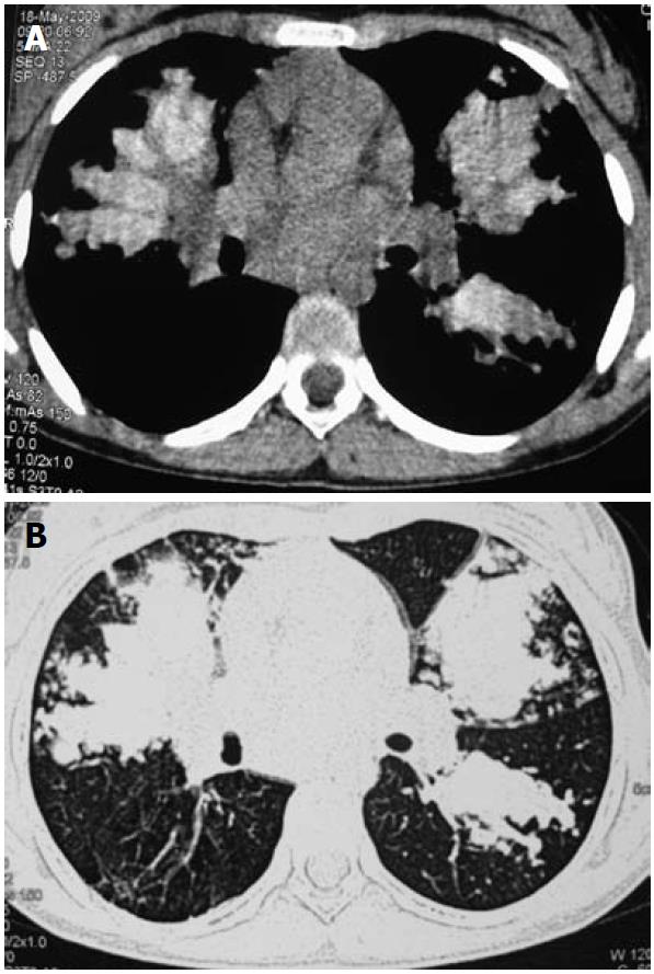
Figure 2 High-resolution computed tomography of the chest showing high-attenuation mucus in a patient with allergic bronchopulmonary aspergillosis (mediastinal window, right panel).
The corresponding lung window is also shown on the left panel.
- Citation: Agarwal R. Allergic bronchopulmonary aspergillosis: Lessons for the busy radiologist. World J Radiol 2011; 3(7): 178-181
- URL: https://www.wjgnet.com/1949-8470/full/v3/i7/178.htm
- DOI: https://dx.doi.org/10.4329/wjr.v3.i7.178


