Copyright
©2011 Baishideng Publishing Group Co.
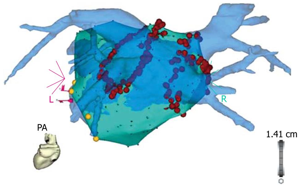
Figure 1 The blue 3D anatomical shell of the left atrium and the pulmonary veins, as acquired by pre-procedural computed tomography, is merged with the grey anatomical shell that was constructed with electro-anatomical mapping during the procedure (CARTO merge).
Note the red ablation tags which mark the circumferential ablation lesions around the pulmonary vein ostia.
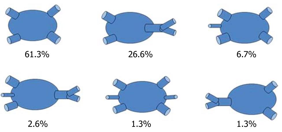
Figure 2 Variants of the left atrium and pulmonary vein-anatomy.
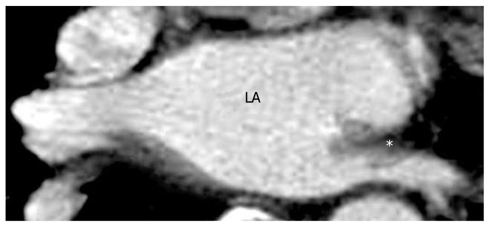
Figure 3 Axial multidetector computed tomography image of the area around a pulmonary vein stenosis (*) into the left inferior pulmonary vein in a 66-year-old male patient with dyspnea and chest discomfort 3 mo after pulmonary vein ablation.
LA: Left atrium.
- Citation: Sohns C, Vollmann D, Luethje L, Dorenkamp M, Seegers J, Schmitto JD, Zabel M, Obenauer S. MDCT in the diagnostic algorithm in patients with symptomatic atrial fibrillation. World J Radiol 2011; 3(2): 41-46
- URL: https://www.wjgnet.com/1949-8470/full/v3/i2/41.htm
- DOI: https://dx.doi.org/10.4329/wjr.v3.i2.41


