Copyright
©2011 Baishideng Publishing Group Co.
World J Radiol. Nov 28, 2011; 3(11): 273-278
Published online Nov 28, 2011. doi: 10.4329/wjr.v3.i11.273
Published online Nov 28, 2011. doi: 10.4329/wjr.v3.i11.273
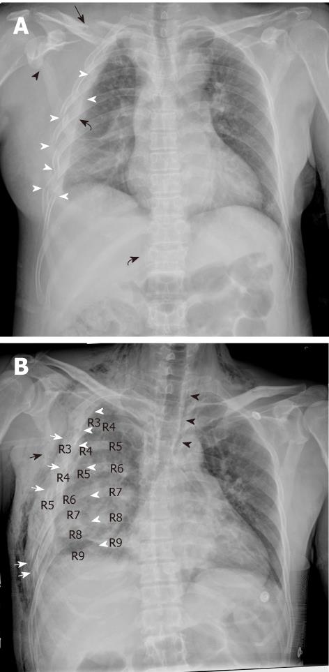
Figure 1 An antero-posterior chest digital radiograph of patients who suffered crush injury in the Sichuan earthquake.
A: Chest digital radiography (CDR) shows the right-sided 2-8 rib fractures at lateral sites (white arrow head), and non-rib thoracic fractures including clavicle (black arrow), scapula (black arrow head) and T12 vertebral body (sinistrad black curved arrow) in a 56-year-old woman after 4 d. Right-sided hemothorax (dextrad black curved arrow) and parenchymal contusion are noted; B: CDR shows thoracic cage asymmetry due to right-sided 3-7 rib fractures and severe dislocations at posterior sites (white arrow head) and lateral sites (white arrow) causing flail chest in a 51-year-old man after 3 d. Right-sided 8 and 9 rib fractures at posterior sites (white arrow head), scapula fracture (black arrow), hemothorax, pneumomediastium (black arrow head) and parenchymal contusion are shown. Note the widespread subcutaneous emphysema.
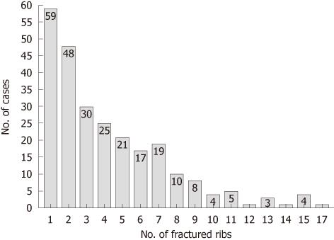
Figure 2 Bar chart shows that the number of fractured ribs per patient ranged from 1 to 17 with a median of 3.
There were 1074 fractured ribs.
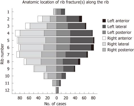
Figure 3 An interactive stack bar shows that most of the rib fractures were distributed from the 3rd through to the 8th ribs, and the vast majority of rib fractures were found in posterior and lateral locations.
- Citation: Dong ZH, Shao H, Chen TW, Chu ZG, Deng W, Tang SS, Chen J, Yang ZG. Digital radiography of crush thoracic trauma in the Sichuan earthquake. World J Radiol 2011; 3(11): 273-278
- URL: https://www.wjgnet.com/1949-8470/full/v3/i11/273.htm
- DOI: https://dx.doi.org/10.4329/wjr.v3.i11.273


