Copyright
©2011 Baishideng Publishing Group Co.
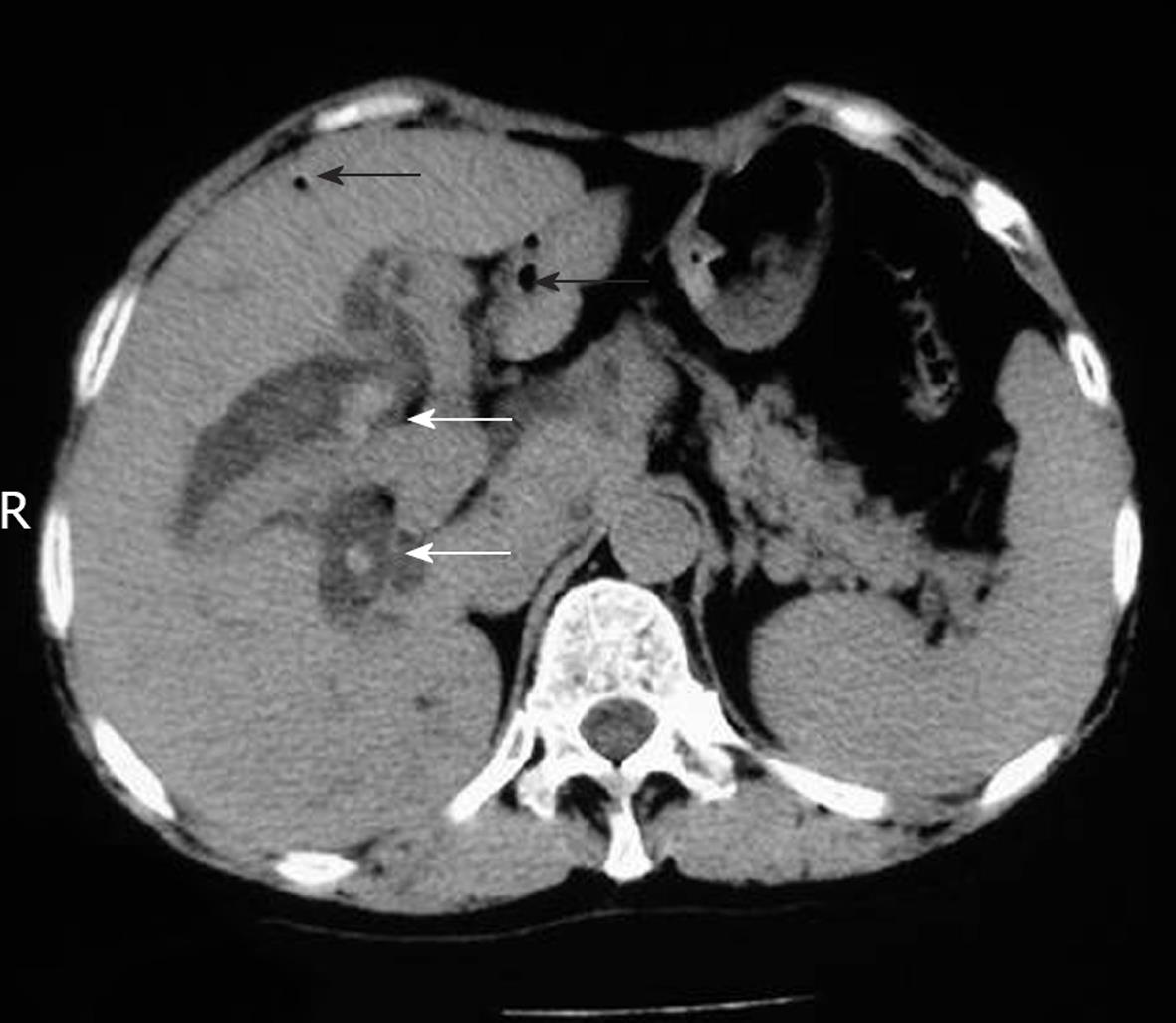
Figure 1 Computed tomography showing air (arrows) and stones (white arrows) in distended bile ducts.
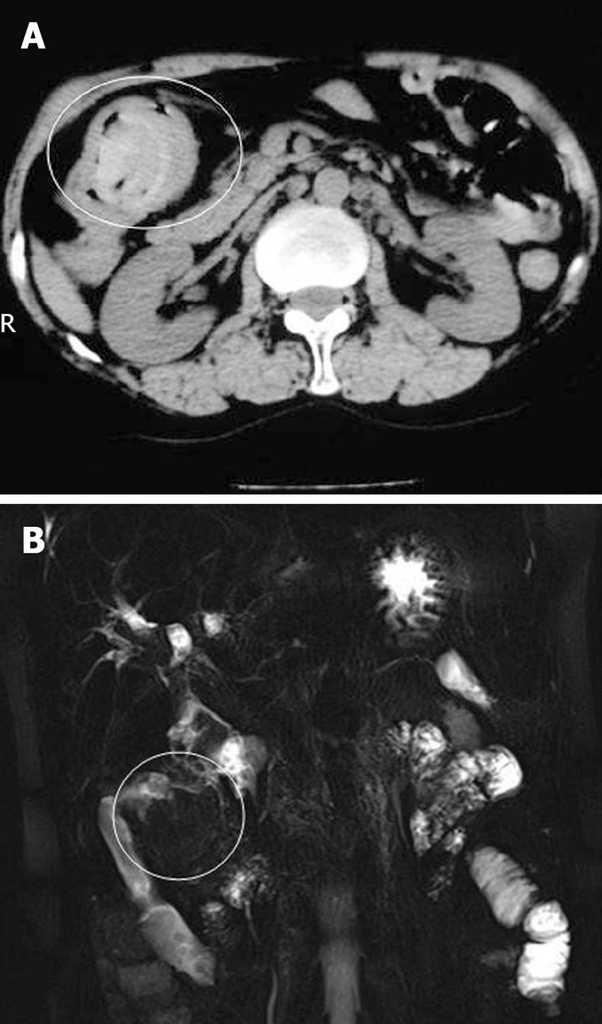
Figure 2 A mass or filling defect in the intestine (white circle).
A: Computed tompograph image; B: Magnetic resonance image.
- Citation: Suo T, Song LJ, Tong SX. Gallstone in jejunal limb with jejunocolonic fistula 10 years after Roux-en-Y choledochojejunostomy. World J Radiol 2011; 3(1): 38-40
- URL: https://www.wjgnet.com/1949-8470/full/v3/i1/38.htm
- DOI: https://dx.doi.org/10.4329/wjr.v3.i1.38


