Copyright
©2010 Baishideng Publishing Group Co.
World J Radiol. Jul 28, 2010; 2(7): 280-282
Published online Jul 28, 2010. doi: 10.4329/wjr.v2.i7.280
Published online Jul 28, 2010. doi: 10.4329/wjr.v2.i7.280
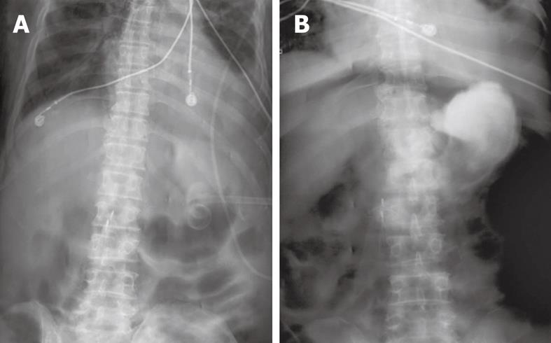
Figure 1 Abdominal imaging.
A: Abdominal X-ray following initial percutaneous endoscopic gastrostomy (PEG) tube placement shows no evidence of free air in the peritoneum; B: Gastrograffin study done on day 14 following PEG tube placement shows free air in the peritoneum without extravasation of contrast into the peritoneum.
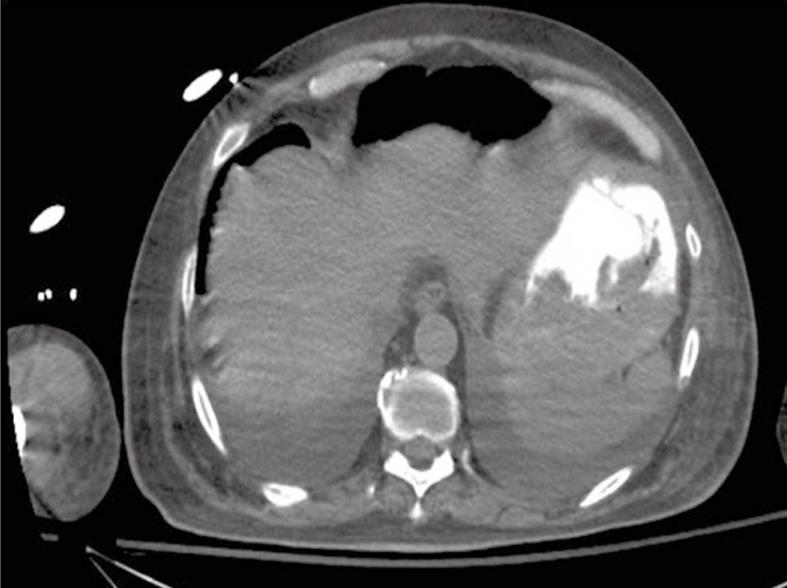
Figure 2 Computed tomography of the abdomen after recurrence of abdominal distension confirming the presence of pneumoperitoneum.
-
Citation: Vijayakrishnan R, Adhikari D, Anand CP. Recurrent tense pneumoperitoneum due to air influx
via abdominal wall stoma of a PEG tube. World J Radiol 2010; 2(7): 280-282 - URL: https://www.wjgnet.com/1949-8470/full/v2/i7/280.htm
- DOI: https://dx.doi.org/10.4329/wjr.v2.i7.280


