Copyright
©2010 Baishideng Publishing Group Co.
World J Radiol. Jul 28, 2010; 2(7): 269-279
Published online Jul 28, 2010. doi: 10.4329/wjr.v2.i7.269
Published online Jul 28, 2010. doi: 10.4329/wjr.v2.i7.269
Figure 1 Lazy-snapping graph-cut identification of the significant biliary structures.
A: Original magnetic resonance cholangiopancreatography thin slice image; B: Interactive identification of foreground and background, with the corresponding object boundaries identified by the algorithm; C: Silhouette of the identified objects.
Figure 2 Dynamic thresholding through histogram analysis to identify significant biliary structures.
A: Original projection-type magnetic resonance cholangiopancreatography image; B: Intensity histogram of the image; C: Resulting thresholded image, rescaled to a 0-255 intensity level.
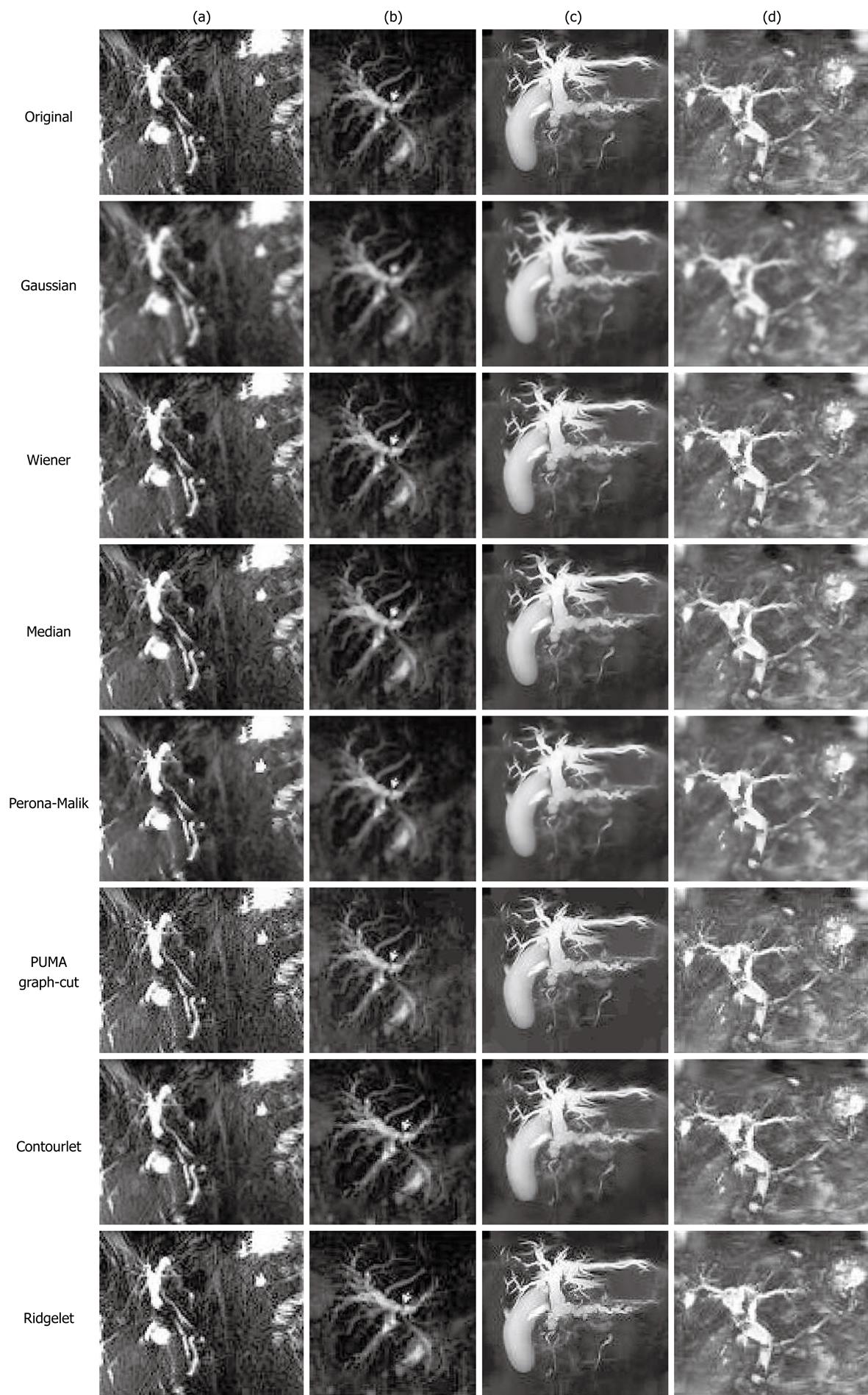
Figure 3 Denoising results for images with heavy tissue background.
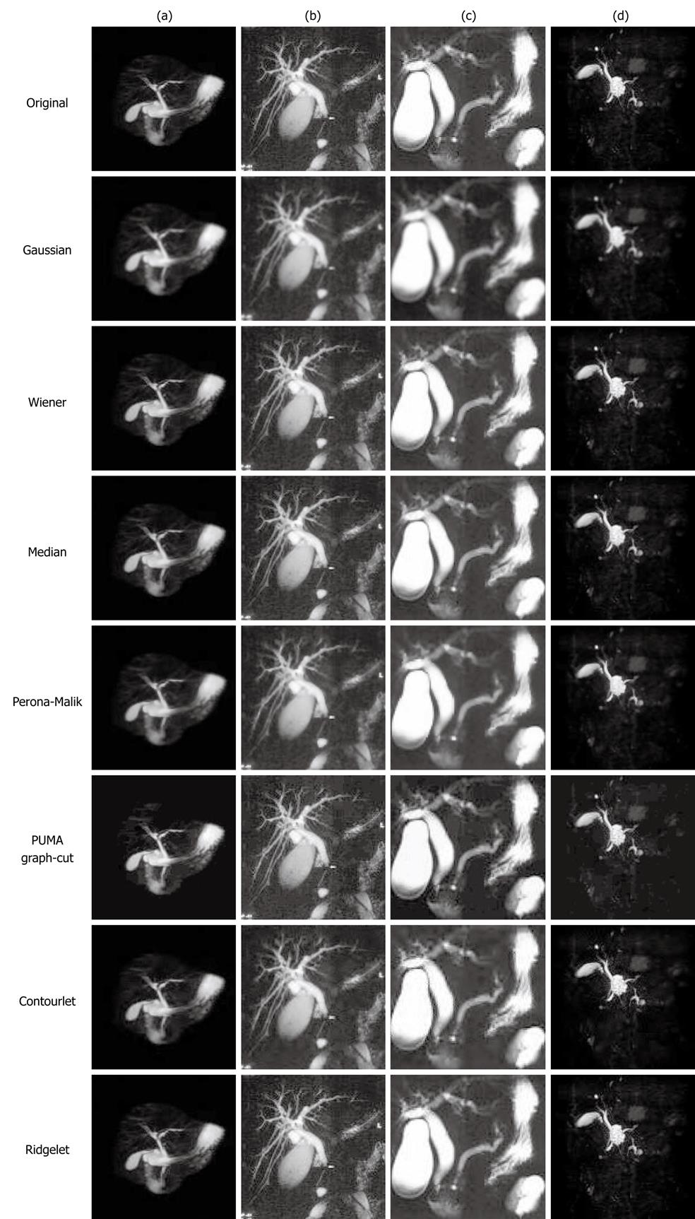
Figure 4 Denoising results for images with multiple objects.
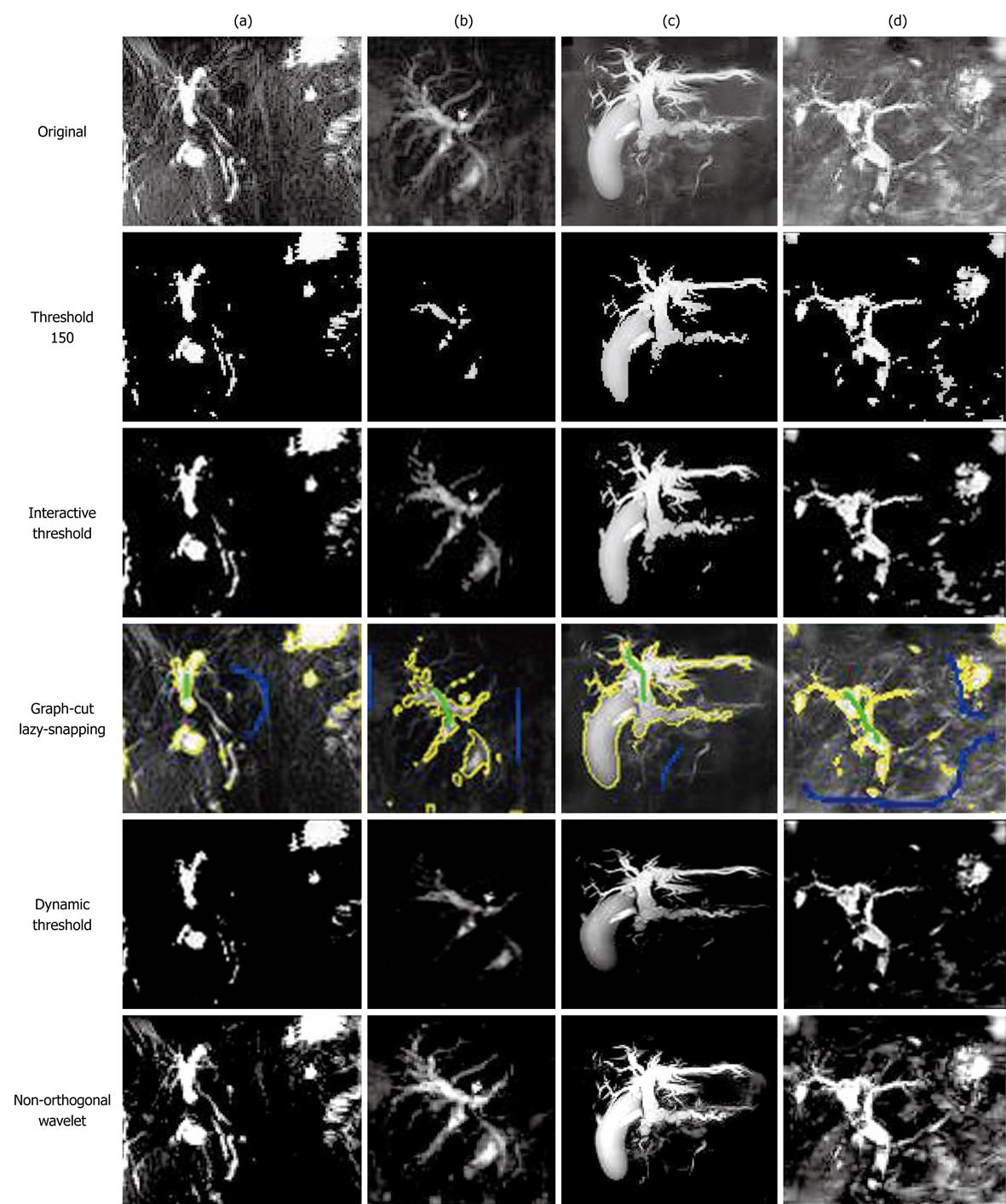
Figure 5 Structure extraction for images with heavy tissue background.
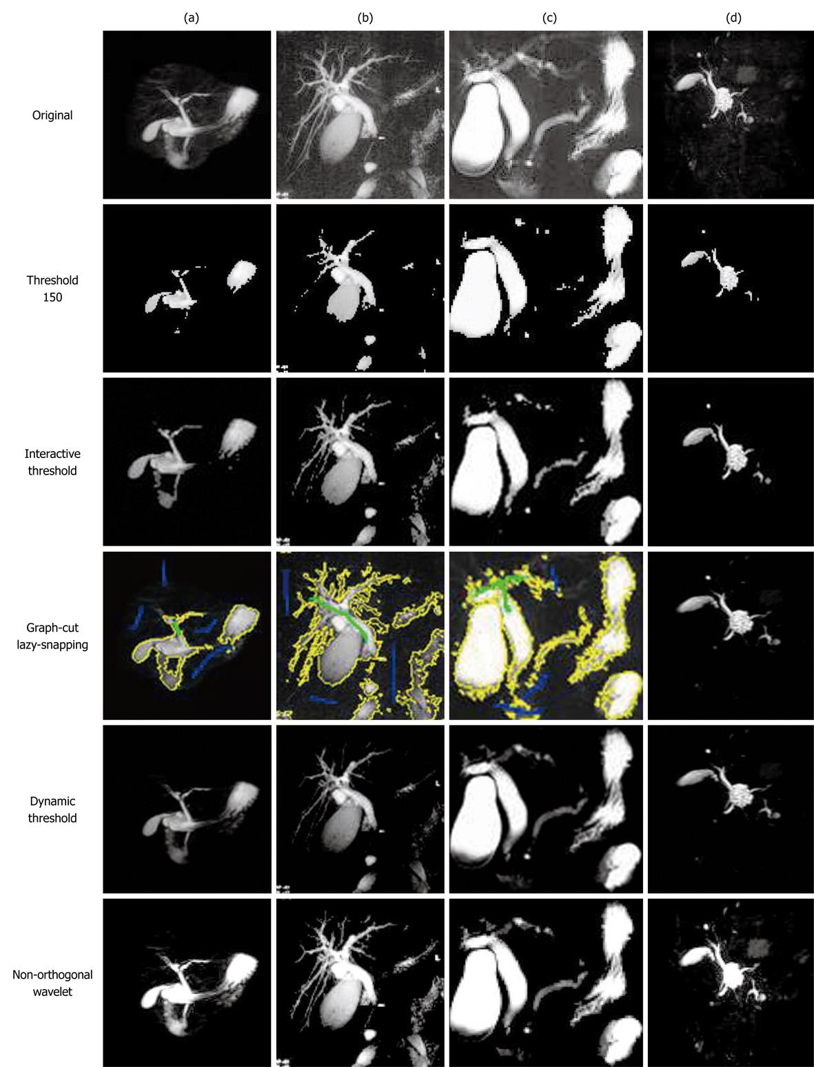
Figure 6 Structure extraction for images with multiple objects.
- Citation: Logeswaran R. Magnetic resonance cholangiopancreatography image enhancement for automatic disease detection. World J Radiol 2010; 2(7): 269-279
- URL: https://www.wjgnet.com/1949-8470/full/v2/i7/269.htm
- DOI: https://dx.doi.org/10.4329/wjr.v2.i7.269


