Copyright
©2010 Baishideng Publishing Group Co.
World J Radiol. Mar 28, 2010; 2(3): 109-112
Published online Mar 28, 2010. doi: 10.4329/wjr.v2.i3.109
Published online Mar 28, 2010. doi: 10.4329/wjr.v2.i3.109
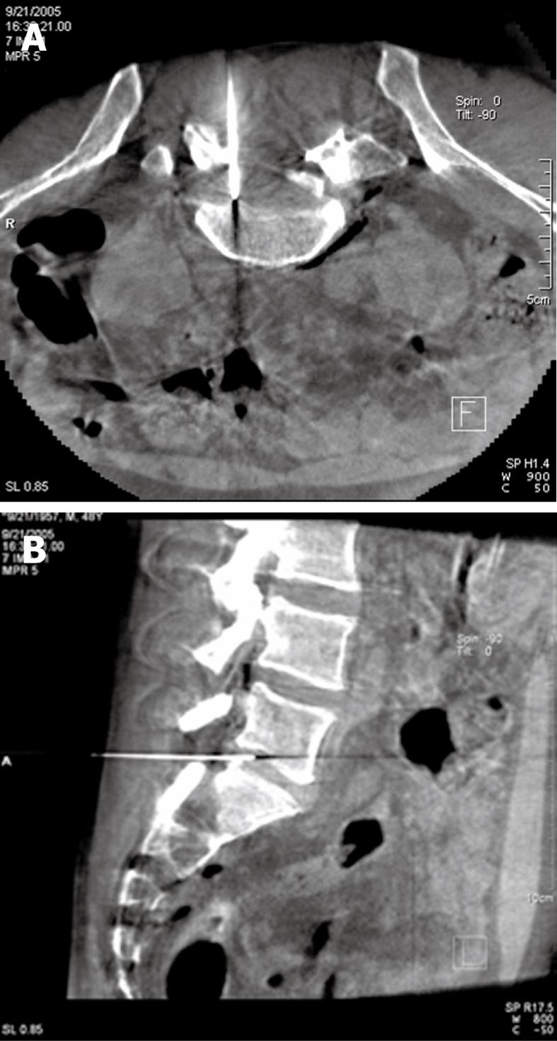
Figure 1 The placement of the needle tip measured accurately by Dyna-CT reconstruction under rotary digital subtraction angiography (DSA).
A: The needle advanced into the disc through the inner margin of the facet joint and the needle tip was placed in the hernia under Dyna-CT cross-sectional reconstruction; B: The needle tip advanced into the disc through the posterior-lateral route under Dyna-CT sagittal reconstruction.
-
Citation: Lu W, Li YH, He XF. Treatment of large lumbar disc herniation with percutaneous ozone injection
via the posterior-lateral route and inner margin of the facet joint. World J Radiol 2010; 2(3): 109-112 - URL: https://www.wjgnet.com/1949-8470/full/v2/i3/109.htm
- DOI: https://dx.doi.org/10.4329/wjr.v2.i3.109


