Copyright
©2010 Baishideng.
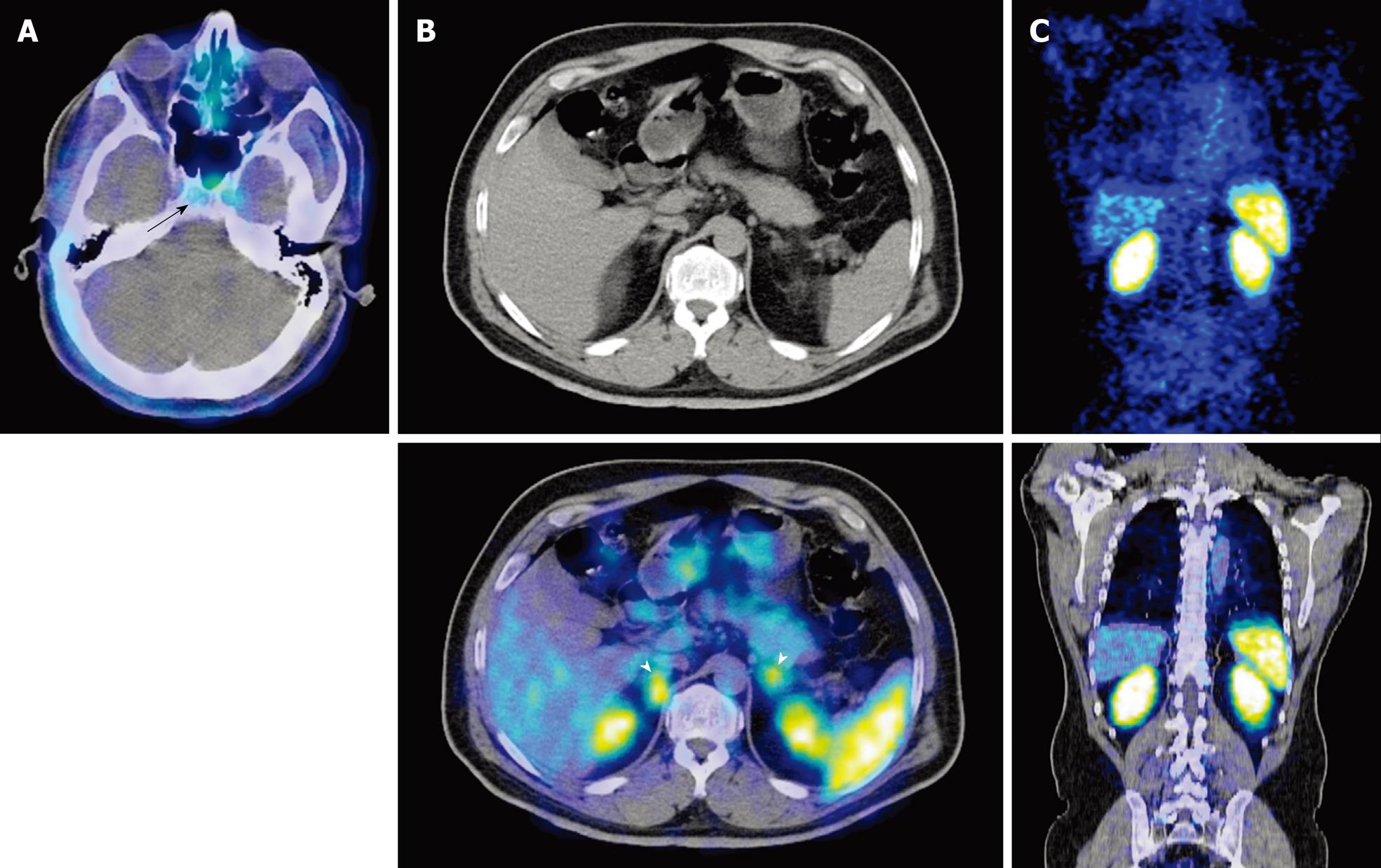
Figure 1 Examples of physiological areas of uptake in somatostatin receptor scintigraphy.
A: Axial positron emission tomography (PET)/computed tomography (CT) of the base of skull demonstrates avid uptake in the pituitary fossa (black arrow) corresponding to physiological uptake in the pituitary gland; B: Axial CT and PET/CT of the abdomen demonstrates physiological uptake in the adrenals glands (white arrowheads); C: Coronal PET and PET/CT sections demonstrate avid tracer uptake in the kidneys and spleen.
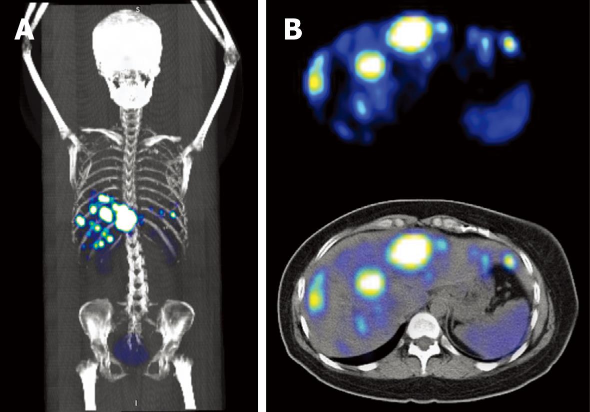
Figure 2 Metastatic pancreatic neuroendocrine carcinoma with multiple liver lesions.
A: Coronal 3D reconstruction demonstrates multiple tracer avid lesions sited predominantly in the right hypochondrium; B: Axial PET and PET/CT of the liver shows multiple somatostatin receptor rich lesions in the liver.
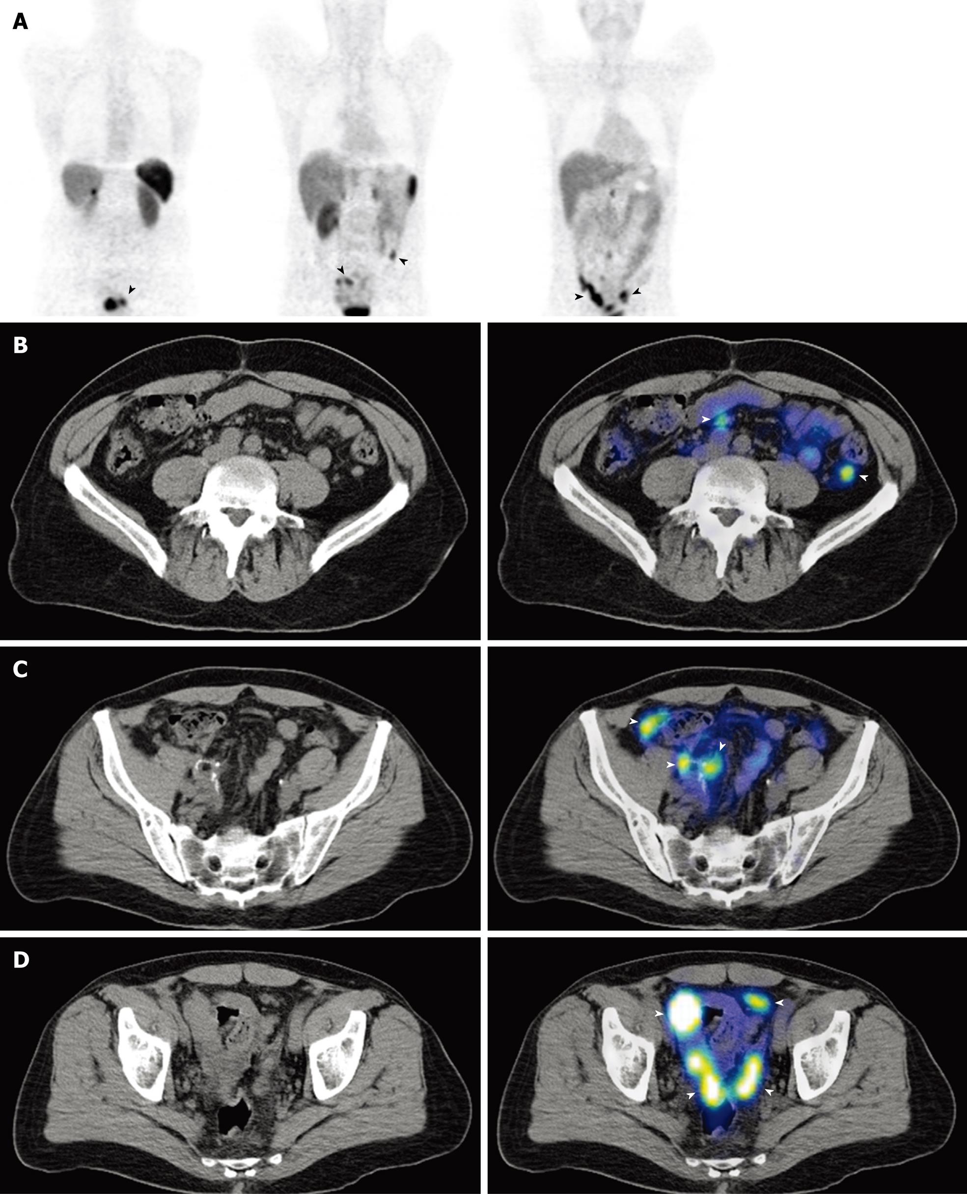
Figure 3 Metastatic appendiceal neuroendocrine carcinoma with multiple peritoneal deposits.
A: Coronal PET images demonstrate multiple tracer avid foci projected over the abdomen and pelvis (black arrowheads); B-D: Axial CT and PET/CT sections of the abdomen show multiple somatostatin receptor rich peritoneal deposits (white arrowheads).
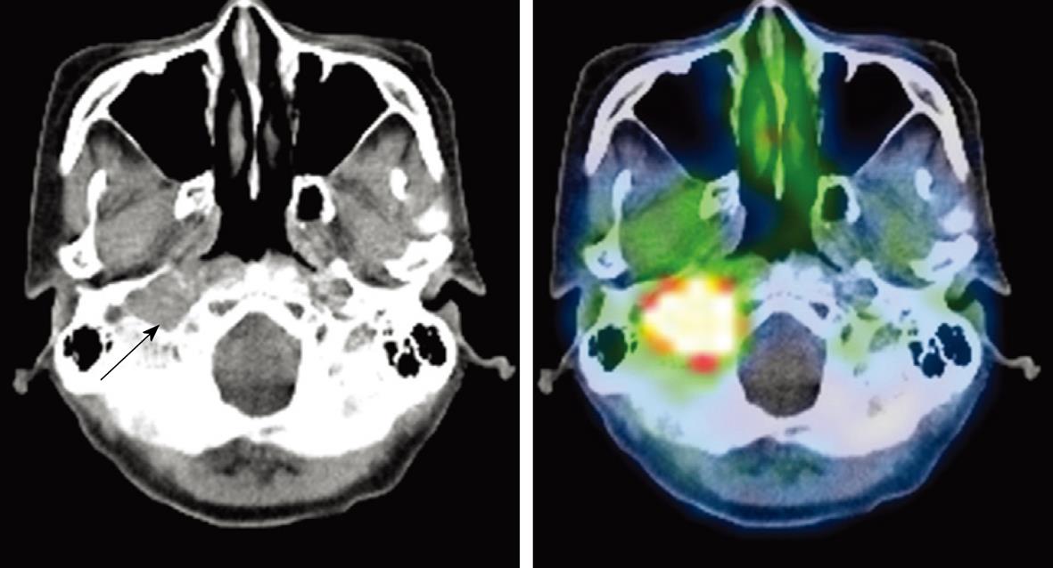
Figure 4 Malignant paraganglioma at the right jugular foramen.
Axial CT and PET/CT of the base of skull. Soft tissue mass in the right jugular foramen showing intense tracer avidity, compatible with a somatostatin receptor expressing tumor.
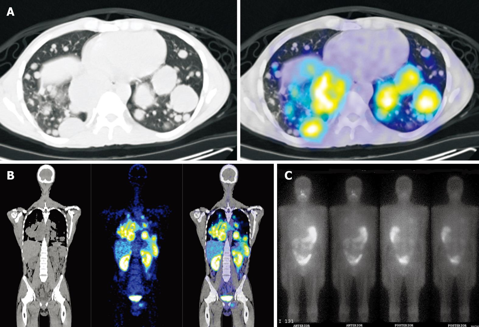
Figure 5 Metastatic non-iodine avid Hurthle cell thyroid carcinoma.
A: Axial CT and PET/CT section of the thorax shows multiple pulmonary masses demonstrating avid tracer uptake; B: Coronal CT, PET and PET/CT images show somatostatin receptor rich pulmonary and mediastinal masses, and another discrete lesion projected over the left pelvis; C: I131 Whole body planar scan of the same patient shows only faint iodine uptake projected over the right upper lobe.
- Citation: Tan EH, Goh SW. Exploring new frontiers in molecular imaging: Emergence of 68Ga PET/CT. World J Radiol 2010; 2(2): 55-67
- URL: https://www.wjgnet.com/1949-8470/full/v2/i2/55.htm
- DOI: https://dx.doi.org/10.4329/wjr.v2.i2.55


