Copyright
©2010 Baishideng Publishing Group Co.
World J Radiol. Oct 28, 2010; 2(10): 410-413
Published online Oct 28, 2010. doi: 10.4329/wjr.v2.i10.410
Published online Oct 28, 2010. doi: 10.4329/wjr.v2.i10.410
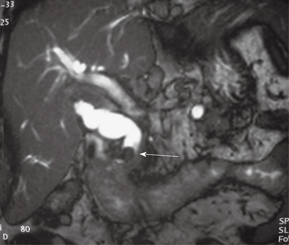
Figure 1 Abdominal magnetic resonance imaging in a sagittal plane demonstrating two stones in common bile duct (arrow) with a diameter of 2.
2 cm and cystic duct, measuring 9 mm and 12 mm respectively, with dilatation of the proximal biliary tree.
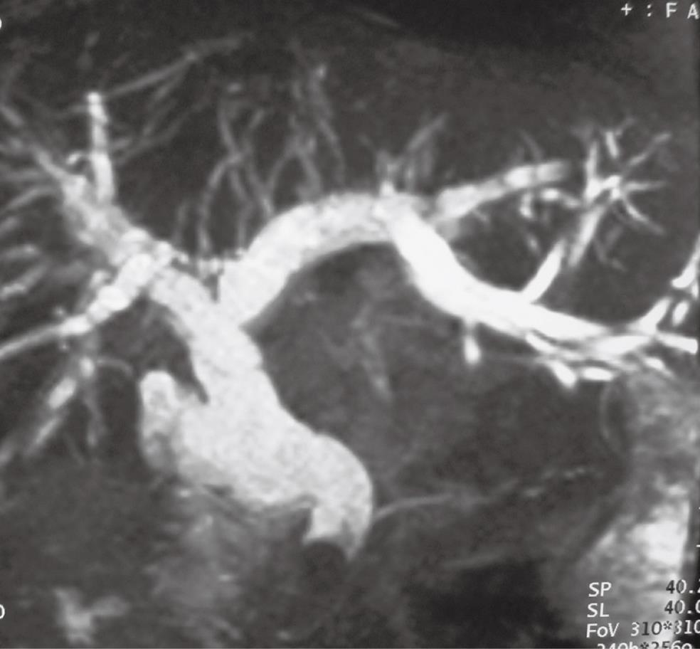
Figure 2 Magnetic retrograde cholangio-pancreatography demonstrating stones in common bile duct and proximal dilatation of the biliary tree.
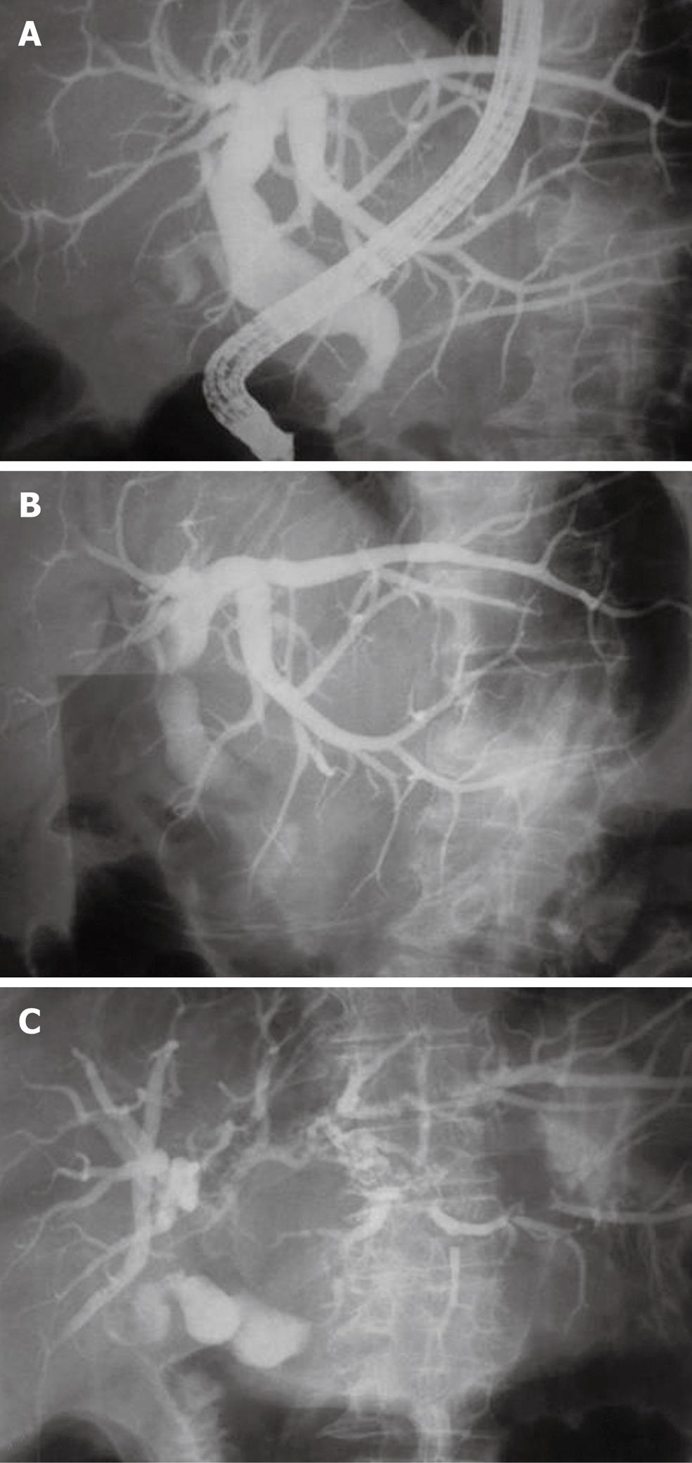
Figure 3 Endoscopic retrograde cholangio-pancreatography showing gradual resolve of dilated biliary tree (A) and indirect signs of Mirizzi syndrome (B and C) when the patient was placed in an anti-Trendelenburg position.
- Citation: Lampropoulos P, Paschalidis N, Marinis A, Rizos S. Mirizzi syndrome type Va: A rare coexistence of double cholecysto-biliary and cholecysto-enteric fistulae. World J Radiol 2010; 2(10): 410-413
- URL: https://www.wjgnet.com/1949-8470/full/v2/i10/410.htm
- DOI: https://dx.doi.org/10.4329/wjr.v2.i10.410


