Copyright
©The Author(s) 2024.
World J Radiol. Jul 28, 2024; 16(7): 247-255
Published online Jul 28, 2024. doi: 10.4329/wjr.v16.i7.247
Published online Jul 28, 2024. doi: 10.4329/wjr.v16.i7.247
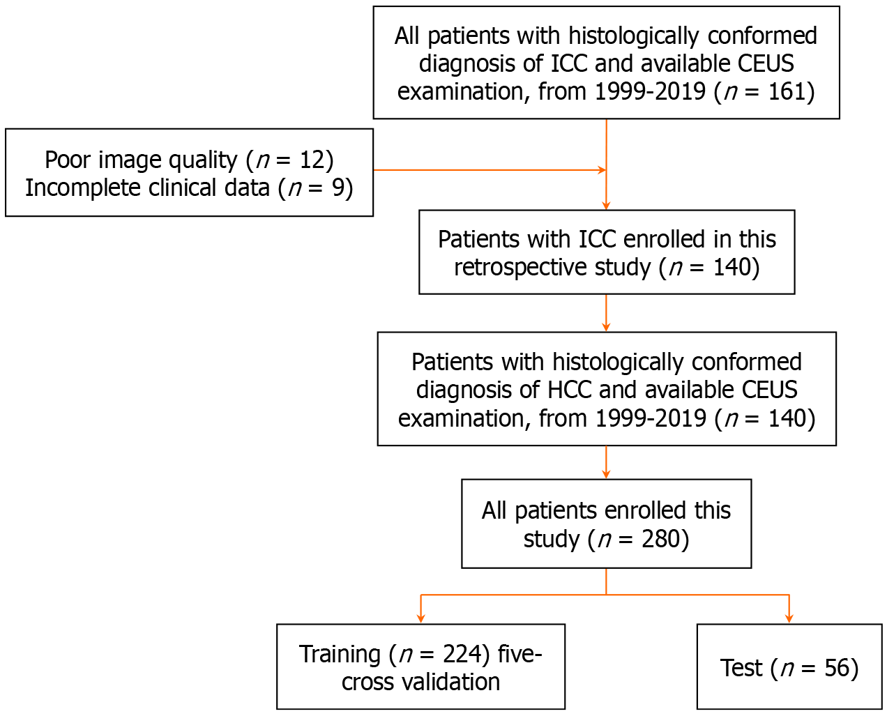
Figure 1 Workflow of necessary steps in current study.
CEUS: Contrast-enhanced ultrasound; HCC: Hepatocellular carcinoma; ICC: Intrahepatic cholangiocarcinoma.
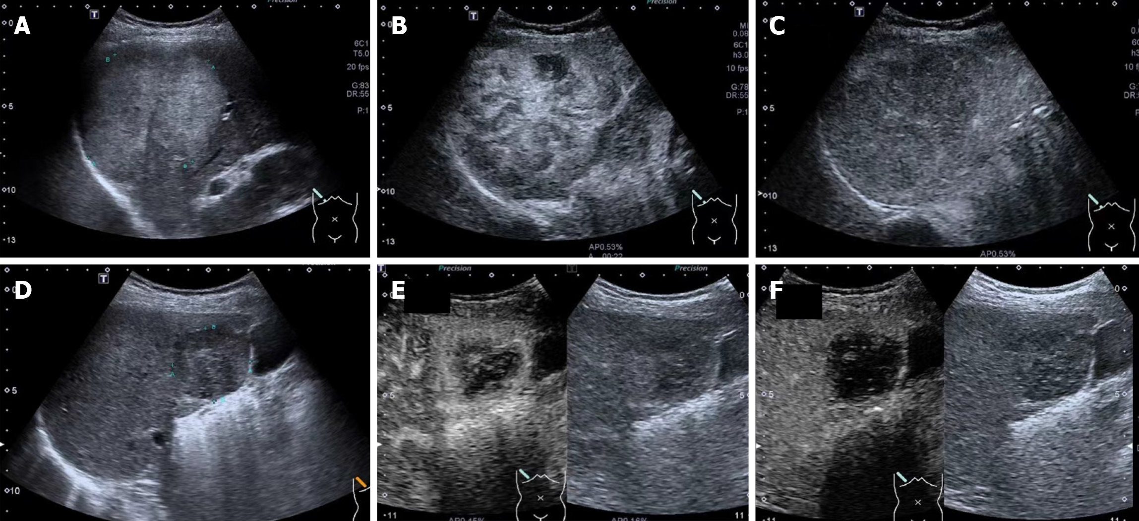
Figure 2 Image acquisition illustration.
A and D: Baseline ultrasound (US) images of two target lesions; B and E: Contrast-enhanced ultrasound (CEUS) images of the target lesions at peak enhancement of arterial phase; C and F: CEUS images of the target lesions at 120 s after injection of contrast agent.
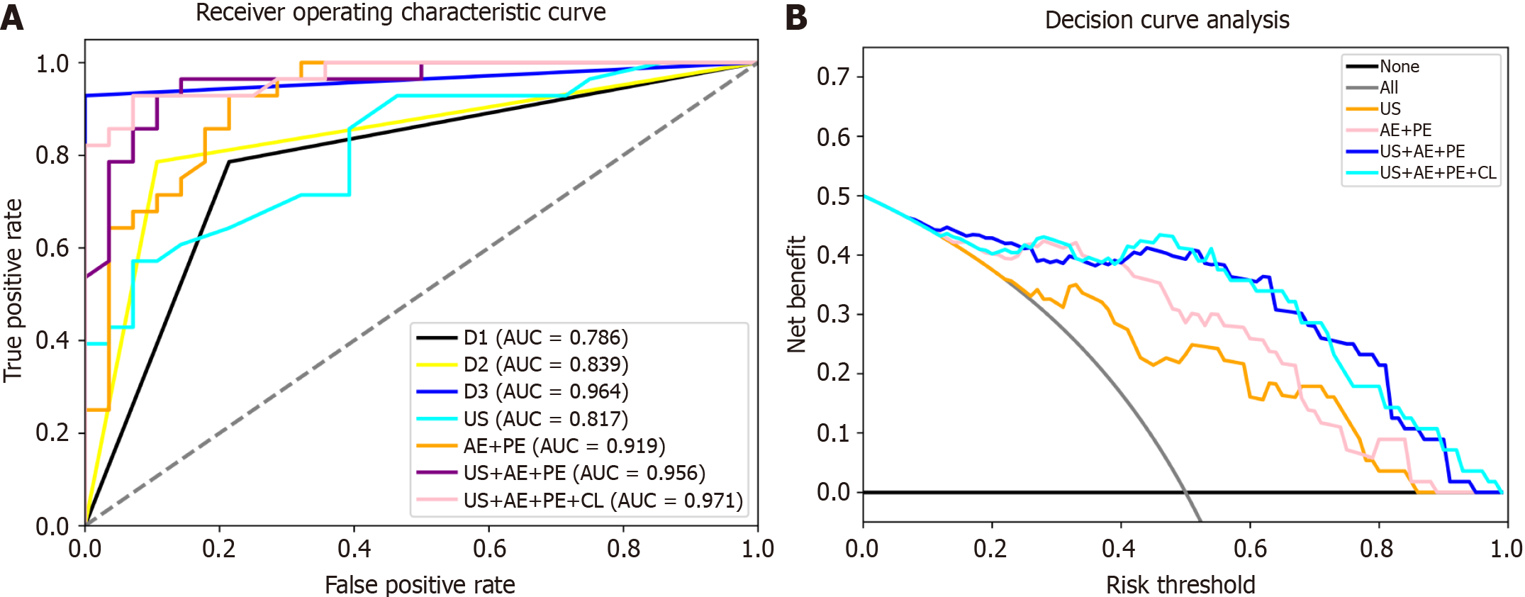
Figure 3 Receiver operating characteristic curves and the decision curve analysis for radiomics models.
A: Receiver operating characteristic curve; B: Decision curve analysis. AE: Arterial enhancement; AUC: Area under the receiver operating characteristic curve; CL: Clinical data; PE: Portal enhancement; US: Ultrasound.
- Citation: Su LY, Xu M, Chen YL, Lin MX, Xie XY. Ultrasomics in liver cancer: Developing a radiomics model for differentiating intrahepatic cholangiocarcinoma from hepatocellular carcinoma using contrast-enhanced ultrasound. World J Radiol 2024; 16(7): 247-255
- URL: https://www.wjgnet.com/1949-8470/full/v16/i7/247.htm
- DOI: https://dx.doi.org/10.4329/wjr.v16.i7.247


