Copyright
©The Author(s) 2020.
Figure 1 Representative axial T2-weighted magnetic resonance image of bilateral carotid arteries with cropped vessel area highlighted (A).
Carotid vessel wall segmentations using convolutional neural networks (B) and expert reader (C). DICE = 0.91.
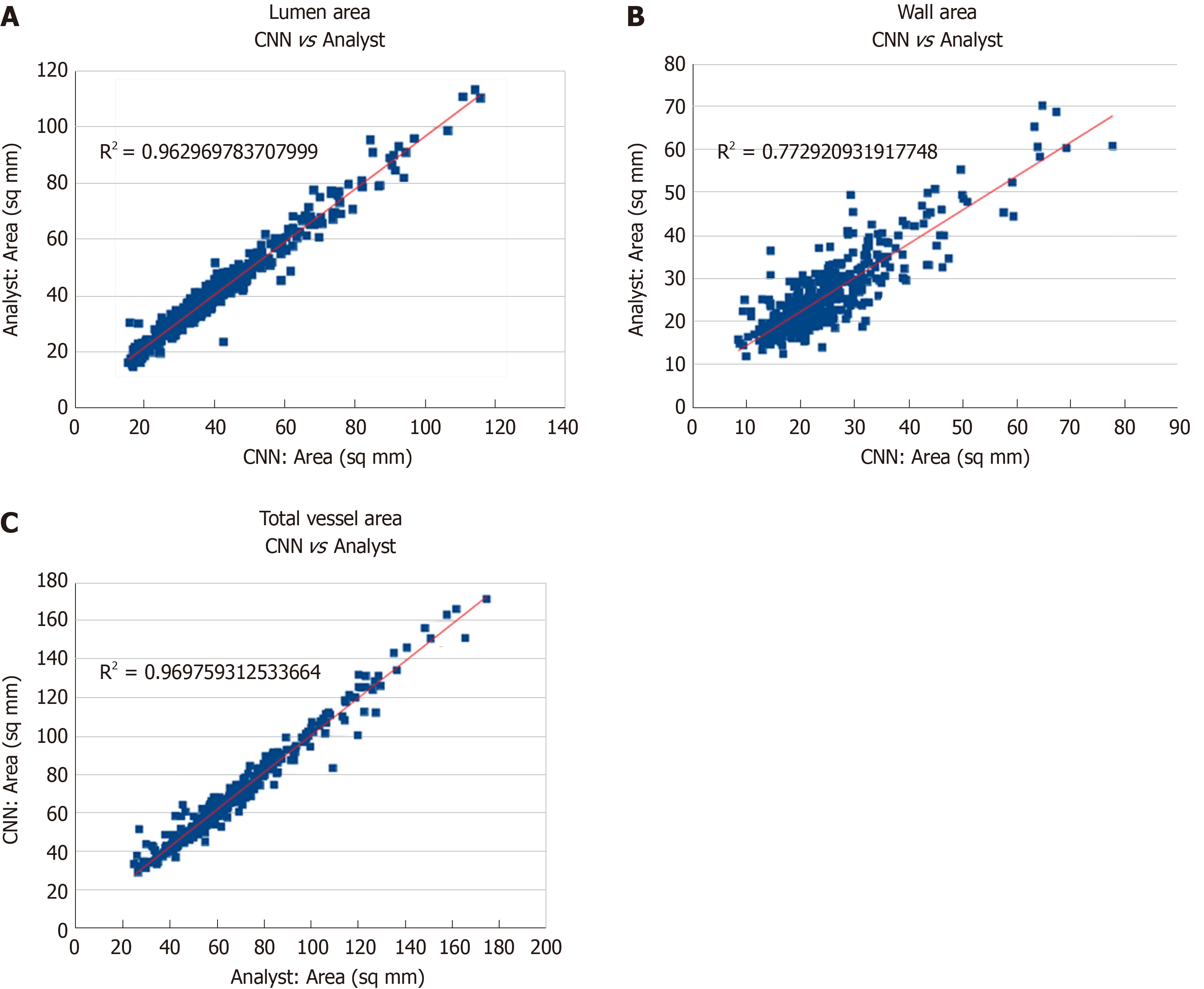
Figure 2 Comparison of measurements between convolutional neural network and an expert reader for lumen area (A), wall area (B), and total vessel area (C).
CNN: Convolutional neural network.
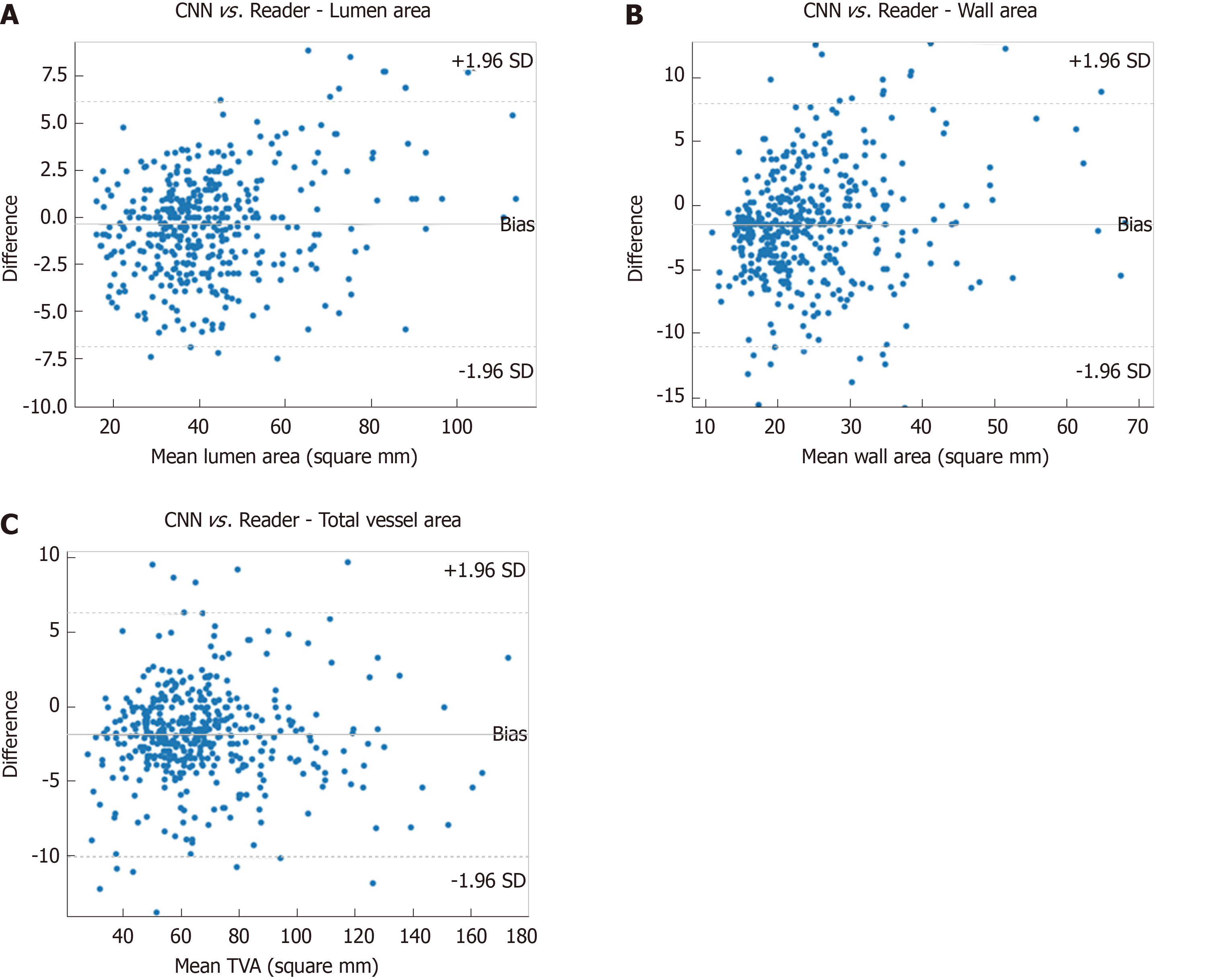
Figure 3 Bland-Altman plots demonstrating agreement of convolutional neural networks and expert reader in assessing lumen area (A), wall area (B), and total vessel area (C).
CNN: Convolutional neural network.
- Citation: Samber DD, Ramachandran S, Sahota A, Naidu S, Pruzan A, Fayad ZA, Mani V. Segmentation of carotid arterial walls using neural networks. World J Radiol 2020; 12(1): 1-9
- URL: https://www.wjgnet.com/1949-8470/full/v12/i1/1.htm
- DOI: https://dx.doi.org/10.4329/wjr.v12.i1.1


