Copyright
©The Author(s) 2019.
World J Radiol. Jul 28, 2019; 11(7): 94-101
Published online Jul 28, 2019. doi: 10.4329/wjr.v11.i7.94
Published online Jul 28, 2019. doi: 10.4329/wjr.v11.i7.94
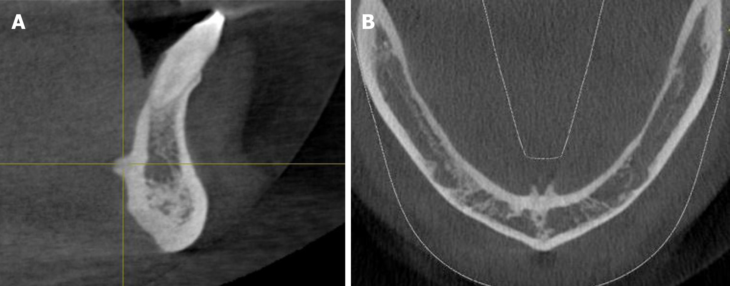
Figure 1 Detection of the genial tubercles pattern in the sagittal and axial views.
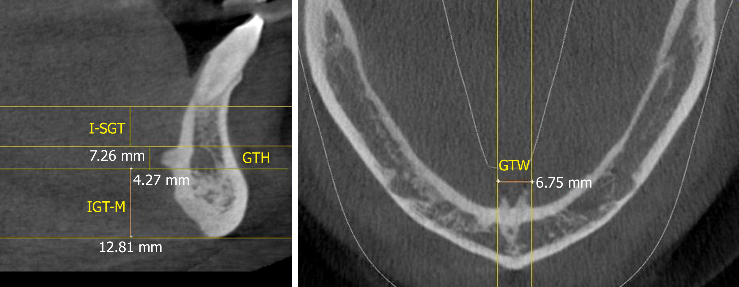
Figure 2 Measurements of the genial tubercle height, distance from the superior border of the genial tubercles to the apex of the lower central incisors and the distance from the inferior border of the genial tubercles to the menton in the sagittal view and genial tubercles width in the axial view.
GTH: Genial tubercles height; GTW: Genial tubercles width; I-SGT: GTs to the lower central incisors; IGT-M: Genial tubercles to the menton.
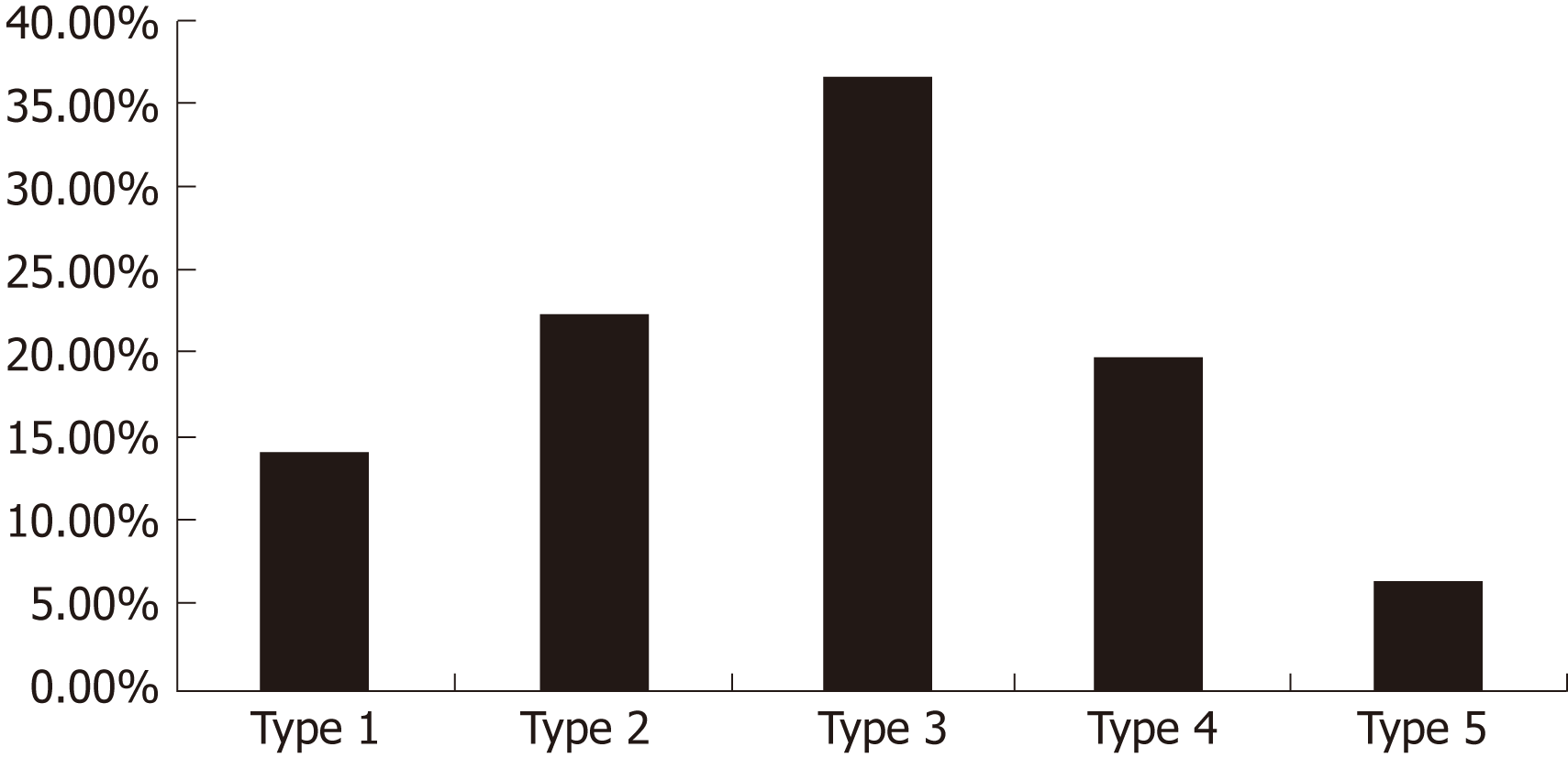
Figure 3 Genial tubercles pattern distribution.
- Citation: Araby YA, Alhirabi AA, Santawy AH. Genial tubercles: Morphological study of the controversial anatomical landmark using cone beam computed tomography. World J Radiol 2019; 11(7): 94-101
- URL: https://www.wjgnet.com/1949-8470/full/v11/i7/94.htm
- DOI: https://dx.doi.org/10.4329/wjr.v11.i7.94


