Copyright
©The Author(s) 2019.
World J Radiol. Jul 28, 2019; 11(7): 102-109
Published online Jul 28, 2019. doi: 10.4329/wjr.v11.i7.102
Published online Jul 28, 2019. doi: 10.4329/wjr.v11.i7.102
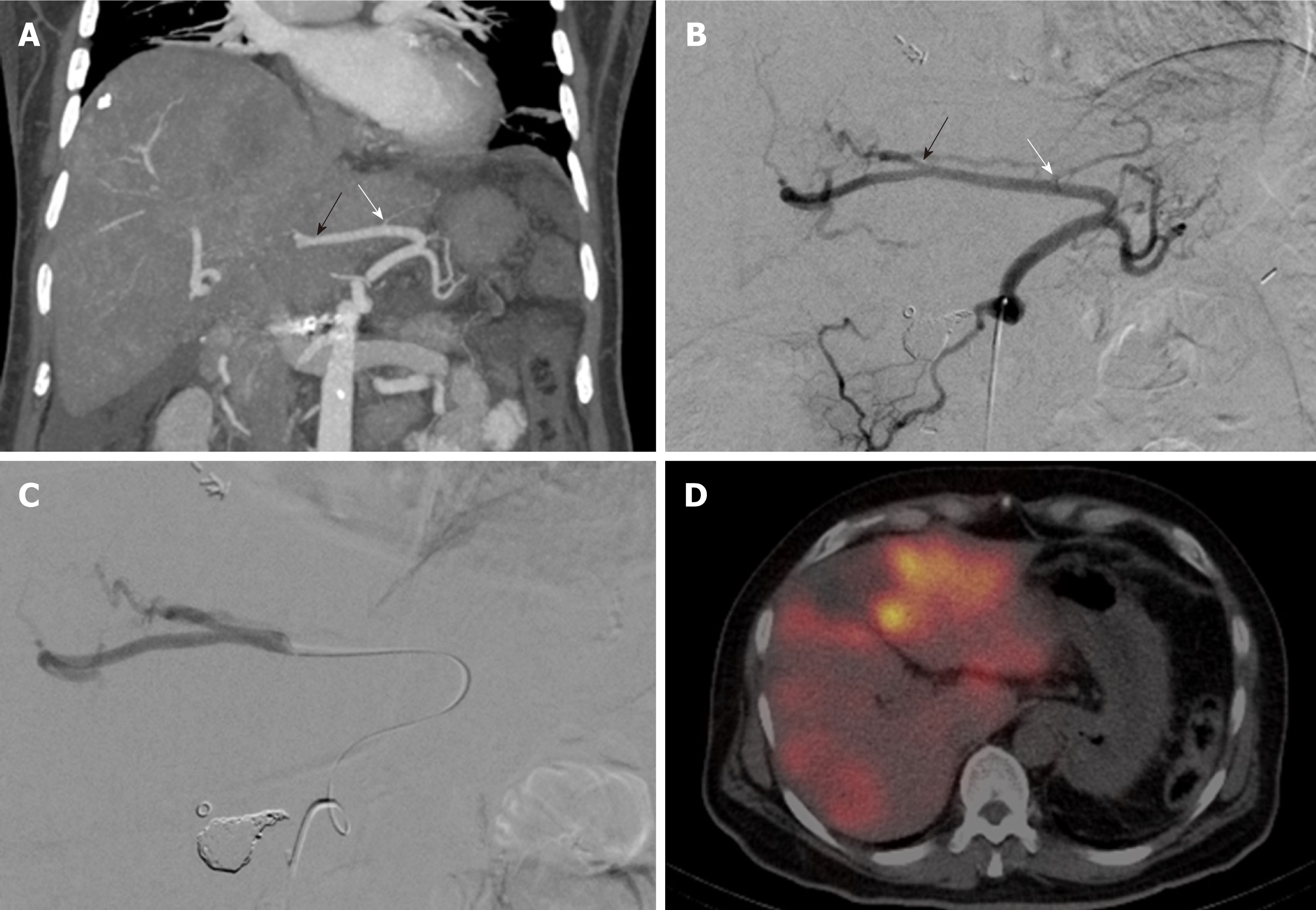
Figure 1 Sample case.
52-year-old patient with an aberrant left hepatic artery originating from the left gastric artery and multifocal colorectal liver metastases in both hepatic lobes. A: Preinterventional computed tomography (CT) angiogram (coronal maximum intensity projection) displaying the distance between the most distal hepatoenteric side branch (white arrow) and the first intrahepatic branch of the aberrant left hepatic artery (LHA) (black arrow); B: Vascular anatomy on the preliminary mapping angiogram. (white arrow: most distal hepatoenteric side branch; black arrow: first intrahepatic branch of the aberrant LHA); C: Catheter position during test injection of technetium 99mTc macro aggregated albumin (99mTc-MAA) (and subsequently also during delivery of the 90Y microspheres); D: Post- 99mTc-MAA SPECT/CT showing good tumoral 99mTc-MAA uptake and no extrahepatic activity.
- Citation: Zimmermann M, Schulze-Hagen M, Pedersoli F, Isfort P, Heinzel A, Kuhl C, Bruners P. Y90-radioembolization via variant hepatic arteries: Is there a relevant risk for non-target embolization? World J Radiol 2019; 11(7): 102-109
- URL: https://www.wjgnet.com/1949-8470/full/v11/i7/102.htm
- DOI: https://dx.doi.org/10.4329/wjr.v11.i7.102


