Copyright
©The Author(s) 2019.
World J Radiol. Dec 28, 2019; 11(12): 144-148
Published online Dec 28, 2019. doi: 10.4329/wjr.v11.i12.144
Published online Dec 28, 2019. doi: 10.4329/wjr.v11.i12.144
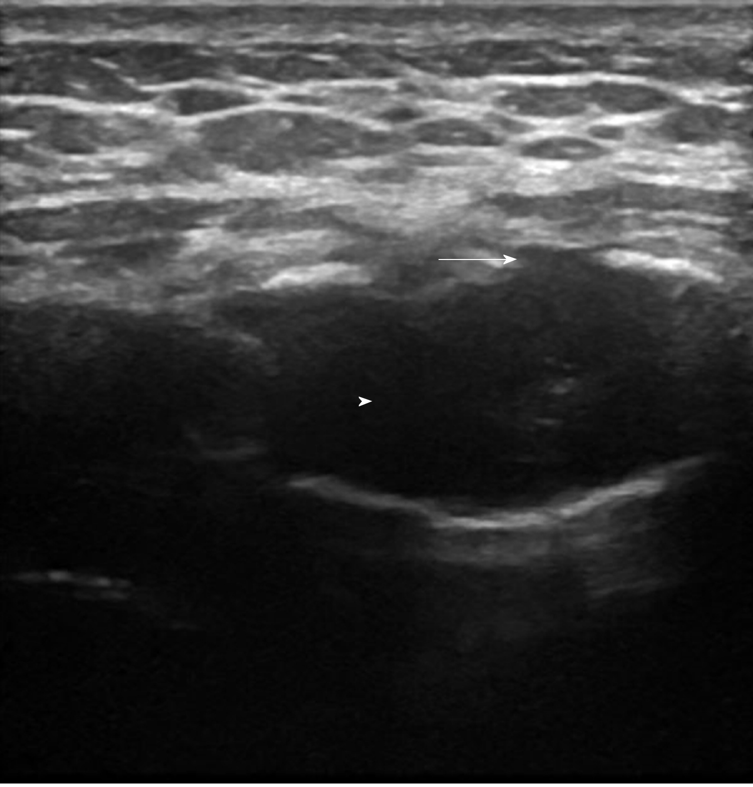
Figure 1 Ultrasound of sternum showing cortical irregularities (arrow) with central hypoechoic area (arrow head).
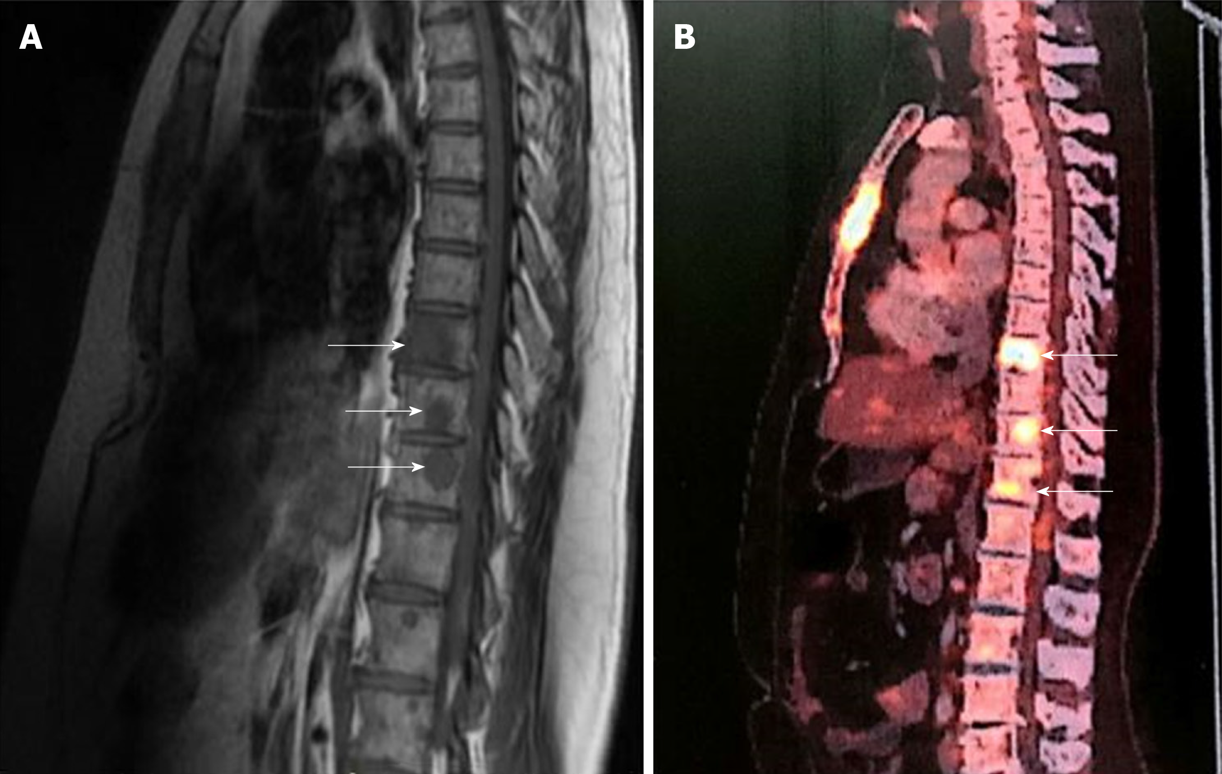
Figure 2 Magnetic resonance imaging and positron emission technology scan.
A: Magnetic resonance imaging showing multiple osteolytic lesions (arrows); B: Positron emission technology scan showing multiple osteolytic lesions with high fluorodeoxyglucose avidity (arrows).
- Citation: Chawla G, Dutt N, Deokar K, Meena VK. Chest pain without a clue-ultrasound to rescue occult multiple myeloma: A case report. World J Radiol 2019; 11(12): 144-148
- URL: https://www.wjgnet.com/1949-8470/full/v11/i12/144.htm
- DOI: https://dx.doi.org/10.4329/wjr.v11.i12.144


