Copyright
©The Author(s) 2018.
World J Radiol. Sep 28, 2018; 10(9): 100-107
Published online Sep 28, 2018. doi: 10.4329/wjr.v10.i9.100
Published online Sep 28, 2018. doi: 10.4329/wjr.v10.i9.100
Figure 1 Phantom experiment.
A: Anatomical image showing cross-section of a water-filled bottle doped with 0.15 mmol/kg of gadolinium (Gd) contrast material. An implantable cardiac defibrillator was placed one inch away from the imaged location; B: The same slice in (A) imaged using the conventional inversion recovery (IR) sequence targeted to null the signal of the doped water in the bottle. The image shows a hyperintensity artifact due to off-resonance signal (arrow); C: The same slice in (B) imaged using the wideband IR sequence, where the hyperintensity artifact was suppressed.
Figure 2 Effect of the improved inversion recovery sequence on removing late gadolinium enhancement hyperintensity artifacts and validation with an electroanatommic map.
A: Conventional late gadolinium enhancement image showing metal hyperintensity artifact (arrows) from the implantable cardioverter defibrillator; B: The same image in (A) acquired with the improved late gadolinium enhancement sequence, which eliminated the artifact and revealed underlying scars (arrows); C: An electroanatommic map of the septal aspect of the right ventricle including the outflow tract. The map is a bipolar voltage map showing low voltage (< 1.5 mV) in the right ventricular outflow tract. The low voltage area (red color) corresponds nicely with the delayed enhancement localized in the septal aspect of the right ventricular outflow tract shown in (B)
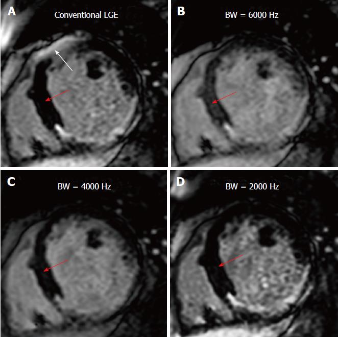
Figure 3 Effect of the inversion recovery bandwidth on myocardial nulling.
A: Conventional late gadolinium enhancement showing a metal hyperintensity artifact (white arrow) mimicking an anterior scar, despite perfect myocardium nulling elsewhere (red arrow); B-D: Using wideband inversion recovery (IR) with different bandwidth (BW). Note that reducing the BW results in improved myocardial nulling (red arrows: the blood-myocardium contrast-to-noise ratio was 20.8, 13.2, 17.1, and 20.1 for conventional IR, and wideband IR with 6000 Hz, 4000 Hz, and 2000 Hz BW, respectively); however, too small BW (< 2000 Hz in this case) results in reappearance of the hyperintensity artifact, similar to that shown in (A).
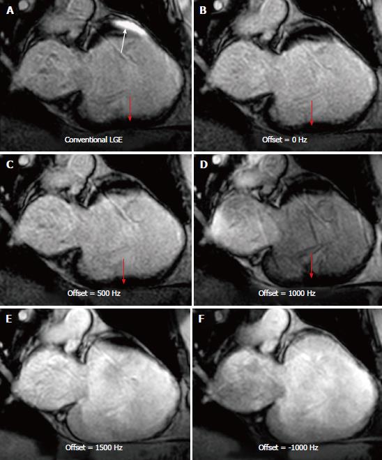
Figure 4 Effects of the inversion recovery frequency offset on myocardial nulling.
A: Conventional late gadolinium enhancement (LGE) showing a metal hyperintensity artifact (white arrow), despite perfect myocardium nulling elsewhere (red arrow); B-F: LGE with wideband inversion recovery (IR). All cases have bandwidth (BW) = 2000 Hz, but different frequency offsets. Note the optimal myocardial nulling in all cases (red arrows in B-D) due to using the small IR BW. However, a too large frequency offset (E and F) results in reappearance of the hyperintensity artifacts that affect most of the myocardial tissues.
- Citation: Ibrahim ESH, Runge M, Stojanovska J, Agarwal P, Ghadimi-Mahani M, Attili A, Chenevert T, den Harder C, Bogun F. Optimized cardiac magnetic resonance imaging inversion recovery sequence for metal artifact reduction and accurate myocardial scar assessment in patients with cardiac implantable electronic devices. World J Radiol 2018; 10(9): 100-107
- URL: https://www.wjgnet.com/1949-8470/full/v10/i9/100.htm
- DOI: https://dx.doi.org/10.4329/wjr.v10.i9.100


