Copyright
©2009 Baishideng Publishing Group Co.
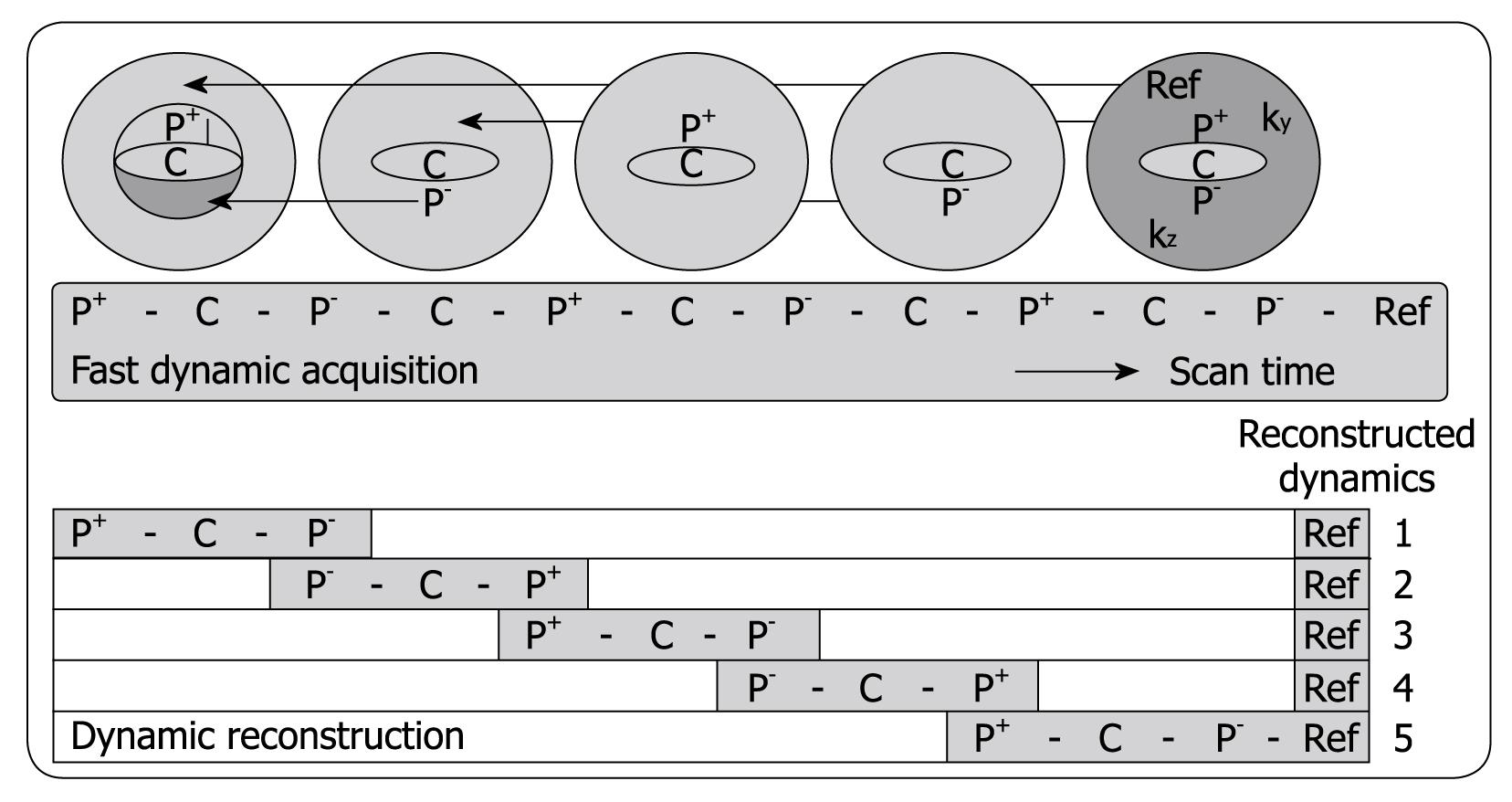
Figure 1 Schematic depiction of the alternating viewsharing technique.
The central ky-kz disk defined by the keyhole percentage is subdivided in 3 regions, P+, C and P-, where P+ and P- cover positive and negative peripheral regions in this central disk and C is the central region as shown. The central region C is acquired in each dynamic scan while regions P+ and P- are shared with subsequent dynamic scans according to an alternating viewsharing scheme: P+-C-P--C-P+-C-P--C-P+-Ref. The P- and P+ parts from subsequent keyhole scans are shared in the reconstruction process.
Figure 2 Liver metastasis in a 68-year-old woman using transverse contrast-enhanced 4D THRIVE.
The reference image using 4D THRIVE in the delayed phase (A) is shown with corresponding automatically calculated Kep map (B). The parametric map shows the liver metastasis (white arrow) as a heterogeneous lesion with ring enhancement. The portal vein is indicated by the white arrowhead.
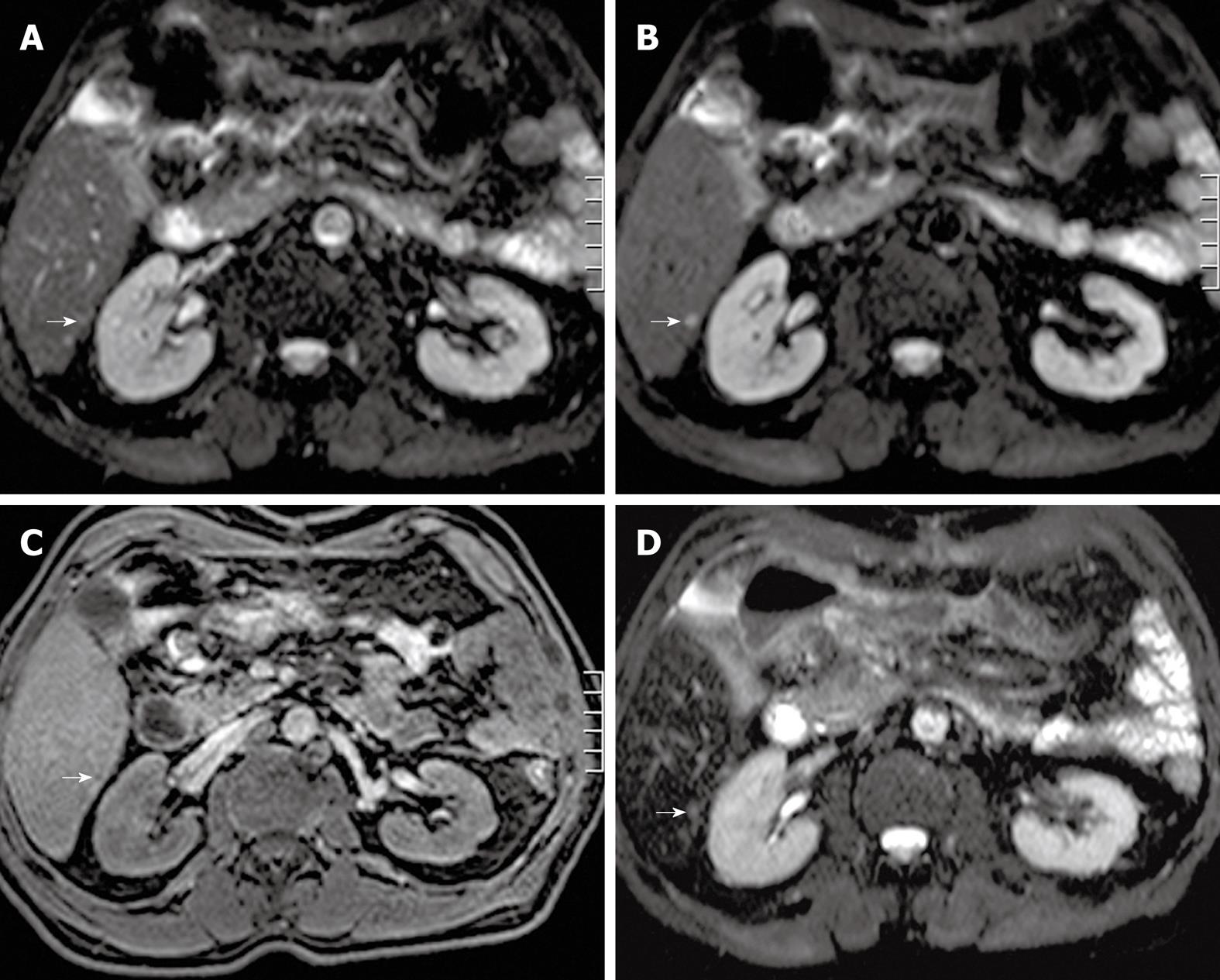
Figure 3 Transverse single-shot spin-echo echo-planar imaging (SS SE-EPI), fat-suppressed T1w 3D GE and SPIO-enhanced T2w TSE (short TE with fat suppression) image in a 62-year-old man.
A: A transverse SS SE-EPI image using b = 0 s/mm2 in a 62-year-old man barely detecting any visible lesion in the area indicated by the white arrow; B: A transverse SS SE-EPI image using b = 10 s/mm2 in the 62-year-old man clearly detecting the liver metastasis (white arrow); C: A transverse fat-suppressed T1w 3D GE image in the portal-venous phase after intravenous injection of SPIO in the 62-year-old man barely detecting the liver metastasis (white arrow); D: A transverse SPIO-enhanced T2w TSE (short TE with fat suppression) image in the 62-year-old man barely detecting the liver metastasis (white arrow). SS SE-EPI: Single-shot spin-echo echo-planar imaging; GE: Gradient echo; T1w: T1-weighted; SPIO: Superparamagnetic iron oxide; TE: Echo time.
- Citation: Coenegrachts K. Magnetic resonance imaging of the liver: New imaging strategies for evaluating focal liver lesions. World J Radiol 2009; 1(1): 72-85
- URL: https://www.wjgnet.com/1949-8470/full/v1/i1/72.htm
- DOI: https://dx.doi.org/10.4329/wjr.v1.i1.72


