Published online Nov 26, 2017. doi: 10.4330/wjc.v9.i11.807
Peer-review started: May 1, 2017
First decision: July 20, 2017
Revised: August 1, 2017
Accepted: August 15, 2017
Article in press: August 16, 2017
Published online: November 26, 2017
Processing time: 206 Days and 20.5 Hours
To compare post-percutaneous coronary intervention (PCI) radial artery occlusion (RAO) incidence between two conventional radial artery compression devices using a novel air-inflation technique.
One hundred consecutive patients post-PCI were randomized 1:1 to Safeguard or TR band compression devices. Post-radial sheath removal, each compression device was inflated with additional 2 mL of air above index bleeding point during air-filled device application and gradually down-titrated accordingly. RAO was defined as absence of Doppler flow signal performed at 24 h and at 6 wk post-PCI. Patients with missing data were excluded. Statistical significance was defined as P < 0.05.
All patients had 6F radial sheath inserted. No significant differences were observed between Safeguard Radial (n = 42) vs TR band (n = 42) in terms of age (63 ± 11 years vs 67 ± 11 years), clinical presentation (electives, n = 18 vs n = 16; acute coronary syndrome, n = 24 vs n = 26) and total procedural heparin (7778 ± 2704 IU vs 7825 ± 2450 IU). RAO incidence was not significantly different between groups at 24 h (2% vs 0%, P = 0.32) and 6 wk (0%, both).
Safeguard Radial and TR band did not demonstrate significant between-group differences in short-term RAO incidence. Lack of evidence of RAO in all post-PCI patients at 6 wk follow-up, regardless of radial compression device indicate advantage of using the novel and pragmatic air-inflation technique. Further work is required to more accurately confirm these findings.
Core tip: Radial artery occlusion (RAO) is a rare but significant complication post-transradial percutaneous coronary intervention (PCI). We found that post-PCI Doppler flow signal-detected RAO incidence was not significantly different between Safeguard Radial and TR band compression devices. However, with the use of a novel air-inflation technique, we observed significantly lower incidence of RAO in all patients regardless radial compression device, in the short-term compared to current literature. Therefore, this novel air-inflation technique may offer a pragmatic and effective solution in reducing RAO incidence.
- Citation: Voon V, AyyazUlHaq M, Cahill C, Mannix K, Ahern C, Hennessy T, SamerArnous, Kiernan T. Randomized study comparing incidence of radial artery occlusion post-percutaneous coronary intervention between two conventional compression devices using a novel air-inflation technique. World J Cardiol 2017; 9(11): 807-812
- URL: https://www.wjgnet.com/1949-8462/full/v9/i11/807.htm
- DOI: https://dx.doi.org/10.4330/wjc.v9.i11.807
Radial artery occlusion (RAO) is an increasingly recognized and significant vascular complication among those observed post-hemostatic compression device application for transradial percutaneous coronary intervention (PCI), the recommended access route in current guidelines[1]. As a consequence of RAO, ipsilateral limb transradial access may be rendered unusable for future procedures. This may be particularly crucial in post-PCI patients, a cohort at higher risk of requiring further coronary angiography, conduit for coronary artery bypass surgery or arterio-venous fistula formation for hemodialysis. Furthermore, ipsilateral ulnar artery access may be unusable due to ischemic limb risk on arterial cannulation.
Several studies have reported rates of RAO from 1%-30%[2-9]. These figures reflect the complex pathophysiology involved in RAO, particularly impaired vascular remodeling and thrombo-inflammatory alterations post-arterial injury. In addition, reports to date have been confounded by multiple external factors. These factors include heterogeneity of study designs, targeted patient populations, parameters for assessing RAO, anticoagulation as well as compression devices and techniques[10].
While several compression bands and techniques tested have demonstrated a modest reduction in RAO, a more pragmatic and effective approach remains to be defined[11]. Therefore, we aimed to prospectively compare incidence of RAO between two conventional hemostatic compression devices (Safeguard Radial and TR band) using a pragmatic and novel air-inflation technique, in patients post-transradial PCI.
Ethics approval was obtained from University Hospital Limerick Ethics committee for our study, which conformed to the principles of the Helsinski Declaration. A total of 107 consecutive patients who had undergone transradial percutaneous coronary intervention at University Hospital Limerick, were screened and eligible patients were recruited into the study. Patients gave written informed consent prior to PCI and were prospectively randomized to either Safeguard Radial or TR band compression device via a pre-specified 1:1 automated randomization. Exclusion criteria were patients less than 18 years old, pregnancy, inability to consent, inability to attend follow-up clinic and difficult radial access requiring femoral access. Patient demographics and angiographic profiles were collected. All patients received dual anti-platelet therapy prior to PCI. Radial artery procedural preparation and management as well as RAO assessment are described below.
After sterile preparation, 1% lidocaine was injected at puncture site. The radial artery was punctured at the anterior wall with a 21-gauge arterial needle through which a 0.018-inch straight floppy tip guidewire (40-cm length) was advanced upon appearance of pulsatile flow. Following this, the needle was withdrawn and a hydrophilic 6F introducer sheath (11-cm length) with dilator length of 16 cm (Prelude, Merit Medical Systems) was inserted over the guidewire into the radial artery. Subsequently, the wire and dilator were removed. According to operator preference, a “radial cocktail” consisting of intra-arterial 100-200 mcg nitroglycerin, 250 mcg verapamil and heparin 2000-4000 IU, was given. All patients had total procedural heparin 70-100 IU/kg given as part of the PCI procedure.
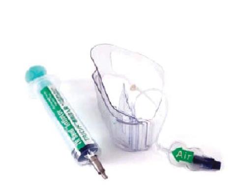
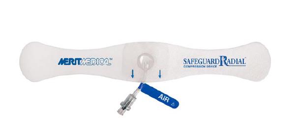
Patients were randomized to either TR band (Terumo Interventional Systems) or Safeguard Radial (Merit Medical) hemostatic compression device groups (Figures 1 and 2). Immediately post-PCI, the radial sheaths were pulled out 4-5 cm and chosen hemostatic compression device band was placed around wrist, with the transparent bladder immediately over the puncture site. We utilized a novel and pragmatic air-inflation technique that involved initial syringe-guided inflation of 2-5 mL of air into device transparent bladder via a cuff-valve system, with careful simultaneous removal of sheath. Continued inflation up to 5-10 mL of air was done to stop bleeding after complete removal of sheath. This was followed by immediate release of air, using similar syringe until bleeding/oozing point, at which an additional 2 mL of air was inflated into device bladder. This is contrary to current non-personalized air-inflation techniques utilizing standard 15 mL and 7 mL of air in TR band and Safeguard respectively, as per manufacturer’s instructions. Subsequent gradual down-titration of air (1 mL of air removed every 30 min) was performed by nursing staff until completion of hemostasis.
Activated clotting time (ACT) is the routine method of choice for monitoring heparin therapy during PCI. At the end of PCI, for all patients, 5 mL of fresh arterial blood sample was obtained in a 5 mL syringe after initially discarding 10 mL of blood from radial sheath prior to sheath removal. The fresh blood was immediately measured for ACT, by using a disposable single-use point-of-care assay. The assay consists of a cuvette containing manufacturer reagents and is measured by the accompanying battery-operated, hand-held device Hemochron Jr Signature + Whole Blood Microcoagulation System (International Technidyne) as per manufacturer’s instructions.
The handheld ultrasonic Doppler signal flow detector 2 MHz (FD1, Huntleigh, Sonicaid) probe was applied above the puncture site of radial artery of resting and extended forearm. RAO was defined as absence of Doppler signal flow. This was performed at 24 h and 6 wk post-PCI by operators blinded to randomization process.
Primary endpoint was RAO at 24 h post-procedure and 6 wk follow-up. Secondary endpoints were bleeding requiring transfusion/surgical intervention, hematoma and pain/numbness at radial access site.
As a pilot study evaluating this technique, exploratory analyses was performed. Continuous normal data were expressed as mean ± SD. Continuous non-normal and categorical variables were expressed as mean (25th, 75th percentile) or frequencies (and percentages). Accordingly, between-group comparisons were compared using unpaired t-testing, Mann-Whitney rank sum test or Pearson chi-square tests. All patients with missing data were excluded from analyses. All analyses were performed using SPSS version 18 statistical software (SPSS Inc, Chicago, IL, United states). P < 0.05 was considered significant (two-tailed significance).
Baseline demographics of patient cohort are presented in Table 1. A total 84 patients were included for analyses after excluding patients who were not eligible (n = 5)/refused (n = 2), had missing data (n = 16). No significant differences were observed between-groups in terms of demographics or procedural profiles (Table 2). Approximately 60% of patients presented with an acute coronary syndrome. All patients had 6F radial sheaths inserted. Despite no significant between-group differences in post-procedural outcomes measures, both Safeguard Radial and TR band groups demonstrated very low incidence of RAO at 24 h (2% vs 0%) and 6 wk (0%, both) (Table 3). No significant differences in secondary outcome measures were observed.
| Variables | Safeguard radial, n = 42 | TR band, n = 42 | P value |
| Age, years | 63.8 ± 10.9 | 66.8 ± 10.8 | 0.21 |
| Gender, male/female ratio | 31/11 | 37/5 | 0.16 |
| BMI, kg/m2 | 29.2 ± 3.9 | 29.0 ± 5.7 | 0.88 |
| Diabetes | 4 (10%) | 8 (19%) | 0.22 |
| CKD | 1 (2%) | 2 (5%) | 0.56 |
| PAD | 0% | 1 (2%) | 0.32 |
| Indication | |||
| Elective | 18 | 16 | 0.88 |
| UA | 6 | 8 | 0.9 |
| NSTEMI | 13 | 8 | 0.7 |
| STEMI | 5 | 10 | 0.6 |
| Variables | Safeguard radial, n = 42 | TR band, n = 42 | P value |
| Pre-procedure | |||
| IR verapamil, % | (86%) | (83%) | 0.77 |
| IR nitroglycerine, % | (31%) | (45%) | 0.18 |
| Procedural | |||
| Heparin, IU | 7778 ± 2704 | 7825 ± 2450 | 0.94 |
| Number of target vessels treated with PCI | |||
| 1 | |||
| 2 | 39 | 39 | 1 |
| ≥ 3 | 3 | 3 | 1 |
| 0 | 0 | 1 | |
| Target vessels | |||
| LM | 1 | 0 | 0.98 |
| LAD/Diagonal | 19 | 22 | 0.87 |
| LCx/OM | 7 | 7 | 0.96 |
| RCA | 18 | 12 | 0.64 |
| IM | 0 | 2 | 0.77 |
| VG | 0 | 2 | 0.77 |
| Fluoroscopy times, min | 15.3 ± 8.4 | 15.0 ± 6.9 | 0.88 |
| Post-procedure | |||
| GP2B3A inhibitor, % | 1 (2%) | 0% | 0.32 |
| ACT ( s ) | 197 ± 38 | 197 ± 47 | 0.97 |
| Variables | Safeguard radial, n = 42 | TR band, n = 42 | P value |
| Bleeding requiring blood transfusion/surgical intervention | 0% | 0% | 1 |
| Hematoma | 7% | 0% | 0.07 |
| Pain/numbness | 2% | 0% | 0.32 |
| Radial artery occlusion at 24 h | 2% | 0% | 0.32 |
| Radial artery occlusion at 6 wk | 0% | 0% | 1 |
This study has demonstrated no significant difference in incidence of short-term RAO between Safeguard and TR band devices. However, we have for the first time demonstrated significantly lower incidence of RAO at 24 h and at 6 wk post-PCI compared to current literature, regardless of type of conventional hemostatic compression device using the novel air-inflation technique. Among the few studies that have reported short-term RAO, some have observed RAO incidence as high as 9.2% at discharge[9]. Pancholy and colleagues reported RAO incidence of 4.4% at 24 h and 3.2% at 30 d using TR band in a cohort using 5F radial sheaths[8]. Dai et al[11] demonstrated that in post-transradial PCI patients, incidence of RAO was at least 11% at 24 h and 10% at 30 d. The study showed that air titration based compression strategy using TR band was superior to non-air titration strategies. However, the study utilized a non-specific, non-personalized method using manufacturer’s instructions.
In our experience, additional 2 mL of air above point of bleeding/oozing provides personalized and adequate temporary patent hemostasis without the need of conventional methods to monitor radial patency. This has been shown despite different surface area of compression bladder of both devices. This magnitude of air may provide sufficient compression on muscle, adipose tissue and artery although impact of higher magnitudes of air remains to be determined. This technique requires confirmation in future studies.
To further support this technique, our study involved a cohort presenting predominantly with acute coronary syndrome, a more prothrombotic state, compared to previous studies. Only 29.7% of transradial PCI-treated patients presented with acute coronary syndrome in a study by Rathore and colleagues[9]. The study demonstrated a higher incidence of RAO as aforementioned with manufacturer’s technique of compression device air inflation. However, several techniques to measure RAO were used and only 50% of patients had hydrophilic radial sheaths compared to our study. Some may argue that lack of sheath hydrophilicity may account for such results.
Furthermore, sheath size has also been a regarded as a contributing factor to RAO. Larger diameter sheaths have been reported to have increase RAO incidence[12-14]. This effect was not observed in our study which used 6F sheaths in all patients who required PCI. Despite that, further studies involving improved imaging modalities are required to more accurately characterize vessel to sheath ratio. This is because the higher prothrombotic effects due to possible oversized sheaths may be offset by heparin therapy that all patients received in our study.
Heparin itself has been shown to reduce incidence of RAO. The lack of procedural heparin is an independent predictor of RAO[9]. Rathore and colleagues demonstrated RAO incidence of 24.1% at 4-6 mo follow-up in those without heparin administration. The study showed that in 92% of patients who had transradial PCI with 6F sheaths, RAO incidence was 8.9% at discharge and 5.6% at follow-up in the TR band group, which demonstrated lesser RAO between compression devices compared. Lefvreet al[15] reported 30% RAO with 1000 IU of heparin. This requires further confirmation, particularly at different comparator doses. However, the results of our study again emphasize the impact of the novel air-inflation technique in reducing RAO beyond conventional anticoagulation.
Several limitations were observed during the study. Firstly, we observed a high prevalence of missing data due to procedures performed out-of hours. However, both groups were well matched in baseline demographics to negate group bias effects. Second, as with all exploratory studies, type I error may contribute to the results. Despite our study demonstrating consistent results at discharge and follow-up, this requires further confirmation. Third, Allen’s test was not routinely performed pre-PCI. However, conventional methods of assessment via plethysmography and oximetry have not yielded consistent results due to influence of collaterals from palmar arches and recanalization[6,16]. Lastly, a known confounding factor that was not measured but critical for vascular management, was increased vigilance using our personalized air-inflation strategy to reduce RAO.
In conclusion, Safeguard Radial and TR band did not demonstrate significant between-group differences in short-term RAO incidence. Lack of evidence of RAO in all post-PCI patients at 6 wk follow-up, regardless of radial compression device indicate advantage of using the novel and pragmatic air-inflation technique. Further work is required to more accurately confirm these findings.
Radial artery occlusion is a rare but significant complication post-transradial percutaneous coronary intervention, which is increasing in its use, globally. Therefore, better radial artery compression techniques are required to reduce such complication.
Conventional radial artery compression devices by varying air-inflation techniques have shown different results in reducing the incidence of radial artery occlusion post-percutaneous coronary intervention. These suggest that novel air-inflation techniques using such devices may yield better results in reducing incidence of radial artery occlusion.
The authors have shown a much lower short-term incidence of post-percutaneous coronary intervention radial artery occlusion, compared to current literature, using a novel and pragmatic air-inflation technique in two conventional radial compression devices, Safeguard Radial and TR band.
This pilot study’s methods and results of this study could be used in a larger prospective study aiming to the impact of this novel air-inflation technique with two conventional radial compression devices in different settings of transradial percutaneous coronary intervention.
Radial artery occlusion is a rare but significant complication of transradial percutaneous coronary intervention. Novel and pragmatic radial compression techniques are required to reduce the incidence of such complication.
This is an interesting manuscript about the comparison of post-percutaneous coronary intervention radial artery occlusion incidence between two conventional radial artery compression devices using a novel air-inflation technique, Safeguard Radial and TR band.
Manuscript source: Unsolicited manuscript
Specialty type: Cardiac and cardiovascular systems
Country of origin: Ireland
Peer-review report classification
Grade A (Excellent): 0
Grade B (Very good): B
Grade C (Good): C, C, C
Grade D (Fair): D
Grade E (Poor): 0
P- Reviewer: Lin GM, Nunez-Gil IJ, Sabate M, Said SAM, Ueda H S- Editor: Kong JX L- Editor: A
E- Editor: Zhao LM
| 1. | Kotowycz MA, Dzavík V. Radial artery patency after transradial catheterization. CircCardiovascInterv. 2012;5:127-133. [RCA] [PubMed] [DOI] [Full Text] [Cited by in Crossref: 122] [Cited by in RCA: 129] [Article Influence: 9.9] [Reference Citation Analysis (0)] |
| 2. | Kiemeneij F, Laarman GJ, Odekerken D, Slagboom T, van der Wieken R. A randomized comparison of percutaneous transluminal coronary angioplasty by the radial, brachial and femoral approaches: the access study. J Am CollCardiol. 1997;29:1269-1275. [PubMed] |
| 3. | Stella PR, Kiemeneij F, Laarman GJ, Odekerken D, Slagboom T, van der Wieken R. Incidence and outcome of radial artery occlusion following transradial artery coronary angioplasty. CathetCardiovascDiagn. 1997;40:156-158. [PubMed] |
| 4. | Nagai S, Abe S, Sato T, Hozawa K, Yuki K, Hanashima K, Tomoike H. Ultrasonic assessment of vascular complications in coronary angiography and angioplasty after transradial approach. Am J Cardiol. 1999;83:180-186. [PubMed] |
| 5. | Zhou YJ, Zhao YX, Cao Z, Fu XH, Nie B, Liu YY, Guo YH, Cheng WJ, Jia DA. [Incidence and risk factors of acute radial artery occlusion following transradial percutaneous coronary intervention]. Zhonghua Yi XueZaZhi. 2007;87:1531-1534. [PubMed] |
| 6. | Sanmartin M, Gomez M, Rumoroso JR, Sadaba M, Martinez M, Baz JA, Iniguez A. Interruption of blood flow during compression and radial artery occlusion after transradial catheterization. Catheter CardiovascInterv. 2007;70:185-189. [RCA] [PubMed] [DOI] [Full Text] [Cited by in Crossref: 135] [Cited by in RCA: 142] [Article Influence: 7.9] [Reference Citation Analysis (0)] |
| 7. | Zankl AR, Andrassy M, Volz C, Ivandic B, Krumsdorf U, Katus HA, Blessing E. Radial artery thrombosis following transradial coronary angiography: incidence and rationale for treatment of symptomatic patients with low-molecular-weight heparins. Clin Res Cardiol. 2010;99:841-847. [RCA] [PubMed] [DOI] [Full Text] [Cited by in Crossref: 94] [Cited by in RCA: 102] [Article Influence: 6.8] [Reference Citation Analysis (0)] |
| 8. | Pancholy S, Coppola J, Patel T, Roke-Thomas M. Prevention of radial artery occlusion-patent hemostasis evaluation trial (PROPHET study): a randomized comparison of traditional versus patency documented hemostasis after transradial catheterization. Catheter CardiovascInterv. 2008;72:335-340. [RCA] [PubMed] [DOI] [Full Text] [Cited by in Crossref: 365] [Cited by in RCA: 382] [Article Influence: 22.5] [Reference Citation Analysis (0)] |
| 9. | Rathore S, Stables RH, Pauriah M, Hakeem A, Mills JD, Palmer ND, Perry RA, Morris JL. A randomized comparison of TR band and radistop hemostatic compression devices after transradial coronary intervention. Catheter CardiovascInterv. 2010;76:660-667. [RCA] [PubMed] [DOI] [Full Text] [Cited by in Crossref: 49] [Cited by in RCA: 50] [Article Influence: 3.6] [Reference Citation Analysis (0)] |
| 10. | Wagener JF, Rao SV. Radial artery occlusion after transradial approach to cardiac catheterization. CurrAtheroscler Rep. 2015;17:489. [RCA] [PubMed] [DOI] [Full Text] [Cited by in Crossref: 32] [Cited by in RCA: 34] [Article Influence: 3.4] [Reference Citation Analysis (0)] |
| 11. | Dai N, Xu DC, Hou L, Peng WH, Wei YD, Xu YW. A comparison of 2 devices for radial artery hemostasis after transradial coronary intervention. J CardiovascNurs. 2015;30:192-196. [RCA] [PubMed] [DOI] [Full Text] [Cited by in Crossref: 19] [Cited by in RCA: 27] [Article Influence: 3.0] [Reference Citation Analysis (0)] |
| 12. | Dahm JB, Vogelgesang D, Hummel A, Staudt A, Völzke H, Felix SB. A randomized trial of 5 vs. 6 French transradial percutaneous coronary interventions. Catheter CardiovascInterv. 2002;57:172-176. [RCA] [PubMed] [DOI] [Full Text] [Cited by in Crossref: 123] [Cited by in RCA: 122] [Article Influence: 5.3] [Reference Citation Analysis (0)] |
| 13. | Takeshita S, Asano H, Hata T, Hibi K, Ikari Y, Kan Y, Katsuki T, Kawasaki T, Masutani M, Matsumura T. Comparison of frequency of radial artery occlusion after 4Fr versus 6Fr transradial coronary intervention (from the Novel Angioplasty USIng Coronary Accessor Trial). Am J Cardiol. 2014;113:1986-1989. [RCA] [PubMed] [DOI] [Full Text] [Cited by in Crossref: 25] [Cited by in RCA: 23] [Article Influence: 2.1] [Reference Citation Analysis (0)] |
| 14. | Wu SS, Galani RJ, Bahro A, Moore JA, Burket MW, Cooper CJ. 8 frenchtransradial coronary interventions: clinical outcome and late effects on the radial artery and hand function. J Invasive Cardiol. 2000;12:605-609. [PubMed] |
| 15. | Lefevre T, Thebault B, Spaulding C, Funck F, Chaveau M, Guillard N, Guillard N, Chalet Y, Bellorini M, Guerin F. Radial artery patency after percutaneous left radial artery approach for coronary angiography. The role of heparin. Eur Heart J. 1995;16:293. |
| 16. | Kwan TW, Ratcliffe JA, Chaudhry M, Huang Y, Wong S, Zhou X, Pancholy S, Patel T. Transulnar catheterization in patients with ipsilateral radial artery occlusion. Catheter CardiovascInterv. 2013;82:E849-E855. [RCA] [PubMed] [DOI] [Full Text] [Cited by in Crossref: 25] [Cited by in RCA: 27] [Article Influence: 2.3] [Reference Citation Analysis (0)] |


