Published online Sep 26, 2014. doi: 10.4330/wjc.v6.i9.1038
Revised: April 15, 2014
Accepted: July 18, 2014
Published online: September 26, 2014
Processing time: 226 Days and 6.4 Hours
Usually, cardiac calcifications are observed in aortic and mitral valves, atrio-ventricular plane, mitral annulus, coronary arteries, pericaridium (usually causing constrictive pericarditis) and cardiac masses. Calcifications of atrial walls are unusual findings that can be identified only using imaging with high spatial resolution, such as cardiac magnetic resonance and computed tomography. We report a case of a 43-year-old patient with no history of heart disease that underwent cardiac evaluation for mild dyspnoea. The echocardiogram showed a calcific aortic valve and a hyper-echogenic lesion located in atrio-ventricular plane. The patient was submitted to cardiac magnetic resonance and to computed tomography imaging to better characterize the localization of mass. The clinical features and location of calcified lesion suggest an infective aetiology causing an endocarditis involving the aortic valve, atrio-ventricular plane and left atrium. Although we haven’t data to support a definite and clear diagnosis, the clinical features and location of the calcified lesion suggest an infective aetiology causing an endocarditis involving the aortic valve, atrio-ventricular plane and left atrium. The patient was followed for 12 mo both clinically and by electrocardiogram and echocardiography without worsening of clinical, electrocardiographic and echocardiographic data. Cardiac magnetic resonance imaging and computed tomography are ideal methods for identifying and following over time patients with calcific degeneration in the heart.
Core tip: A patient was submitted to echocardiography, cardiac magnetic resonance and to computed tomography imaging to better characterize a hyper-echogenic lesion located in the atrio-ventricular plane. The clinical features and location of the calcified lesion suggest an infective aetiology causing an endocarditis involving the aortic valve, atrio-ventricular plane and left atrium.
- Citation: Dattilo G, Anfuso C, Casale M, Giugno V, Camarda L, Laganà N, Di Bella G. Calcific left atrium: A rare consequence of endocarditis. World J Cardiol 2014; 6(9): 1038-1040
- URL: https://www.wjgnet.com/1949-8462/full/v6/i9/1038.htm
- DOI: https://dx.doi.org/10.4330/wjc.v6.i9.1038
Calcification can be observed in many cardiac localizations but is particularly rare as a lesion that involves the aortic valve, atrioventricular plane and left atrium.
We report a case of a 43-year-old patient with no history of heart disease who underwent cardiac evaluation for mild dyspnoea. On physical examination he showed only a mild aortic systolic murmur. Blood pressure (130/65 mmHg) and electrocardiogram were normal. The echocardiogram showed an increase of left ventricular (LV) outflow aortic velocity (max velocity 2.2 m/s) due to calcific aortic valve and a hyper-echogenic lesion located in the atrio-ventricular plane. The patient was submitted to cardiac magnetic resonance (CMR) and to computed tomography imaging to better characterize the localization of mass.
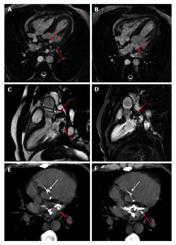
CMR by steady-state free precession sequence showed normal atrial and ventricular dimensions; furthermore hypointense areas located in the left atrium and atrio-ventricular plane (Figure 1, red arrows on panel A-D) with a partial obstruction of superior pulmonary vein (Figure 1, on panel B) were found. A gradient echo T1-weighted image after 10 min of injection of contrast media (delayed contrast enhancement technique) showed a hypointense area in left atrial (LA) suggesting calcium.
Axial images by cardiac computed tomography showed the presence of a mass suggestive of calcium in LA (Figure 1, red arrows on panel E-F), atrioventricular groove and aortic LV outflow (white arrows on panel E-F).
The patient was followed for 12 mo both clinically and by electrocardiogram and echocardiography without worsening of clinical, electrocardiographic and echocardiographic data.
Calcification can be observed in many cardiac localizations[1-7]; particularly, they can be located: (1) valves (usually aortic and mitral valve); (2) atrio-ventricular plane; (3) mitral annulus (usually located in mitral posterior annulus as consequence of a degenerative disorders in the elderly, osteoporosis women, kidney disease); (4) epicardial coronaries; (5) cardiac masses (caseous calcification of the posterior mitral annulus, soft tissue calcified sarcomas, calcified echinococcoccus cysts, cardiac osteocondromas and cardiac calcified amorphous tumors); and (6) in pericaridium (usually causing constrictive pericarditis).
The calcifications of atrial walls are unusual findings that can be identified only using imaging with high spatial resolution, such as cardiac magnetic resonance and computed tomography. Cardiac magnetic resonance imaging and computed tomography, having a high spatial resolution and tissue characterization, are ideal methods for identifying and following over time patients with unusual localization of calcific degeneration in the heart. This case report represents a very rare manifestation of extended endocarditis. Although we haven’t data to support a definite and clear diagnosis, the clinical features and location of the calcified lesion suggest an infective aetiology causing an endocarditis involving the aortic valve, atrio-ventricular plane and left atrium.
A 43-year-old patient with no history of heart disease who underwent cardiac evaluation for mild dyspnoea.
At physical examination there was only a mild aortic systolic murmur.
Cardiac magnetic resonance (CMR) by steady-state free precession sequence showed hypointense areas located in the left atrium and atrio-ventricular plane with a partial obstruction of the superior pulmonary vein and the delayed contrast enhancement technique showed a hypointense area in left atrial (LA) suggesting the presence of calcium. Axial images by cardiac computed tomography showed the presence of a mass suggestive of calcium in LA, atrioventricular groove and aortic left ventricular outflow.
Endocarditis is a serious condition that can endanger patient life, showing itself in different ways.
CMR delayed contrast enhancement technique is based on the use of gradient echo T1-weighted images 10 min after the injection of contrast medium and it is very useful to evaluate the tissue characteristics, particularly in an organ in constant motion like the heart.
This case report not only represents one of the largest extensions of endocarditis described but also shows a lack of correlation between clinical manifestation and clinical symptoms.
The report is interesting, and it is an excellent work.
P- Reviewer: Patanè S, Rostagno C S- Editor: Wen LL L- Editor: O’Neill M E- Editor: Liu SQ
| 1. | Funada A, Kanzaki H, Kanzaki S, Takahama H, Amaki M, Hasegawa T, Yamada N, Kitakaze M. Coconut left atrium. Int J Cardiol. 2012;154:e42-e44. [RCA] [PubMed] [DOI] [Full Text] [Cited by in Crossref: 4] [Cited by in RCA: 3] [Article Influence: 0.2] [Reference Citation Analysis (0)] |
| 2. | Lee WJ, Son CW, Yoon JC, Jo HS, Son JW, Park KH, Lee SH, Shin DG, Hong GR, Park JS. Massive left atrial calcification associated with mitral valve replacement. J Cardiovasc Ultrasound. 2010;18:151-153. [RCA] [PubMed] [DOI] [Full Text] [Full Text (PDF)] [Cited by in Crossref: 10] [Cited by in RCA: 11] [Article Influence: 0.7] [Reference Citation Analysis (0)] |
| 3. | Müller UM, Gielen S, Schuler GC, Gutberlet M. Endocardial calcification of left atrium, tracheobronchopathia osteoplastica, and calcified aortic arch in a patient with dyspnea. Circ Heart Fail. 2008;1:290-292. [RCA] [PubMed] [DOI] [Full Text] [Cited by in Crossref: 4] [Cited by in RCA: 5] [Article Influence: 0.3] [Reference Citation Analysis (0)] |
| 4. | Di Bella G, Masci PG, Ganame J, Dymarkowski S, Bogaert J. Images in cardiovascular medicine. Liquefaction necrosis of mitral annulus calcification: detection and characterization with cardiac magnetic resonance imaging. Circulation. 2008;117:e292-e294. [RCA] [PubMed] [DOI] [Full Text] [Cited by in Crossref: 30] [Cited by in RCA: 31] [Article Influence: 1.8] [Reference Citation Analysis (0)] |
| 5. | Vidal A, Lluberas N, Florio L, Gómez A, Russo D, Agorrody V, Albistur S, Lluberas R. Massive left atrial calcification, tracheobronchopathia osteoplastica and mitral paravalvular leak associated with cardiac rheumatic disease and previous mitral valve replacement. Int J Cardiol. 2013;167:e111-e112. [RCA] [PubMed] [DOI] [Full Text] [Cited by in Crossref: 3] [Cited by in RCA: 4] [Article Influence: 0.3] [Reference Citation Analysis (0)] |
| 6. | Di Bella G, Gaeta M, Pingitore A, Oreto G, Zito C, Minutoli F, Anfuso C, Dattilo G, Lamari A, Coglitore S. Myocardial deformation in acute myocarditis with normal left ventricular wall motion--a cardiac magnetic resonance and 2-dimensional strain echocardiographic study. Circ J. 2010;74:1205-1213. [PubMed] |
| 7. | Di Bella G, Minutoli F, Zito C, Recupero A, Donato R, Carerj S, Coglitore S, Lentini S. Calcified disease of the mitral annulus: a spectrum of an evolving disease. Ann Cardiol Angeiol (Paris). 2011;60:102-104. [PubMed] |


