Copyright
©The Author(s) 2017.
World J Cardiol. Sep 26, 2017; 9(9): 742-748
Published online Sep 26, 2017. doi: 10.4330/wjc.v9.i9.742
Published online Sep 26, 2017. doi: 10.4330/wjc.v9.i9.742
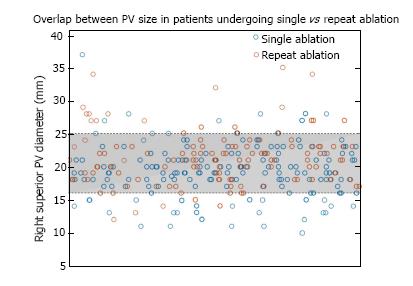
Figure 1 Distribution of right superior pulmonary vein ostial diameter measurements.
There was significant overlap in the distributions of patients with single and repeat procedure, with 80% of all measurements falling between 16 and 25 mm. PV: Pulmonary vein.
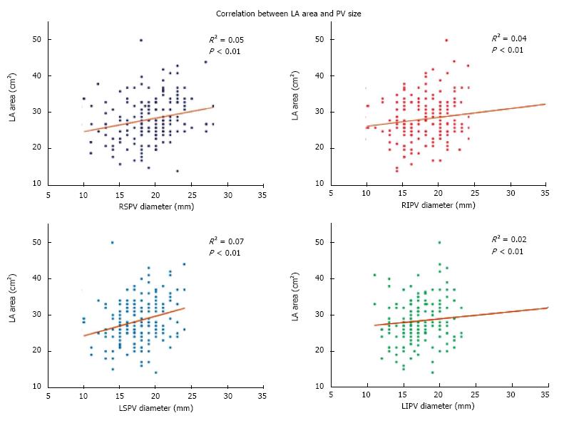
Figure 2 Correlation between pulmonary vein size and left atrial area among all patients in the cohort.
All but the left inferior pulmonary vein (PV) were significantly correlated with left atrial (LA) area, although the correlation coefficients were small. RSPV: Right superior pulmonary vein; RIPV: Right inferior pulmonary vein; LSPV: Left superior pulmonary vein; LIPV: Left inferior pulmonary vein.
- Citation: Desai Y, Levy MR, Iravanian S, Clermont EC, Kelli HM, Eisner RL, El-Chami MF, Leon AR, Delurgio DB, Merchant FM. Clinical and anatomic predictors of need for repeat atrial fibrillation ablation. World J Cardiol 2017; 9(9): 742-748
- URL: https://www.wjgnet.com/1949-8462/full/v9/i9/742.htm
- DOI: https://dx.doi.org/10.4330/wjc.v9.i9.742


