Copyright
©The Author(s) 2017.
World J Cardiol. Sep 26, 2017; 9(9): 742-748
Published online Sep 26, 2017. doi: 10.4330/wjc.v9.i9.742
Published online Sep 26, 2017. doi: 10.4330/wjc.v9.i9.742
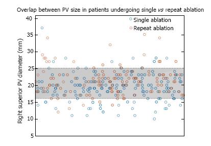
Figure 1 Distribution of right superior pulmonary vein ostial diameter measurements.
There was significant overlap in the distributions of patients with single and repeat procedure, with 80% of all measurements falling between 16 and 25 mm. PV: Pulmonary vein.
- Citation: Desai Y, Levy MR, Iravanian S, Clermont EC, Kelli HM, Eisner RL, El-Chami MF, Leon AR, Delurgio DB, Merchant FM. Clinical and anatomic predictors of need for repeat atrial fibrillation ablation. World J Cardiol 2017; 9(9): 742-748
- URL: https://www.wjgnet.com/1949-8462/full/v9/i9/742.htm
- DOI: https://dx.doi.org/10.4330/wjc.v9.i9.742


